Unsupervised Deep Learning for Limited-Angle STEM-EDX Tomography -- Application to 3D Chemical Analysis of Phase-Change Memory Devices
Pith reviewed 2026-06-27 11:40 UTC · model grok-4.3
The pith
Multi-channel deep image prior with TV regularization reconstructs 3D elemental maps from limited-angle STEM-EDX data without external priors.
A machine-rendered reading of the paper's core claim, the machinery that carries it, and where it could break.
Core claim
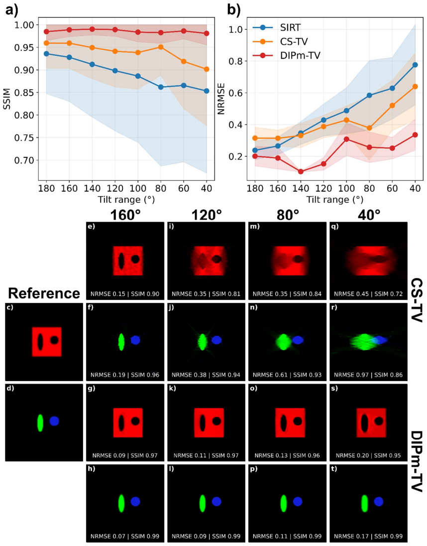
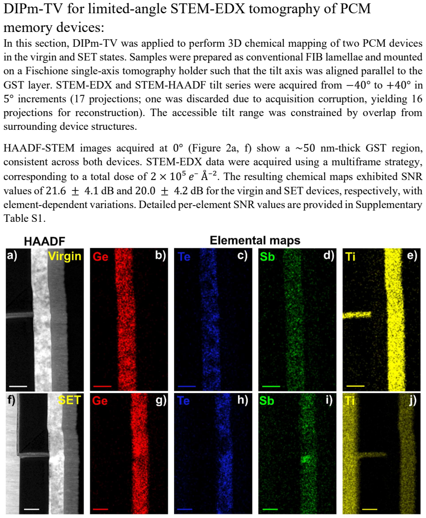
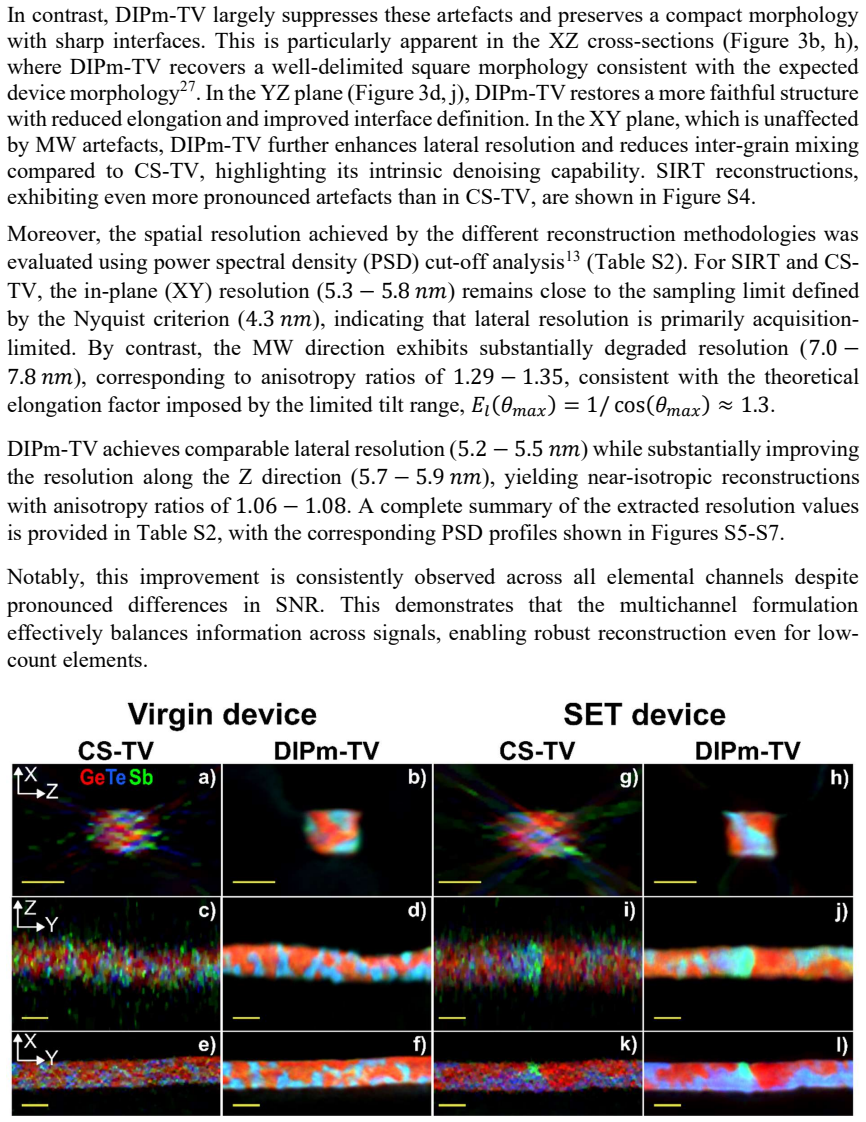
The multi-channel DIPm-TV formulation compensates for severe missing-wedge artefacts corresponding to approximately 100° of missing angular range under moderate noise, enabling voxel-by-voxel elemental reconstruction of GST devices from -40° to +40° tilt data acquired at 5° steps and a dose of 2.0×10^5 e⁻/Ų, without external structural priors such as HAADF imaging, and yields near-isotropic resolution while outperforming SIRT and compressed sensing.
What carries the argument
Multi-channel Deep Image Prior with total variation regularization (DIPm-TV), which represents the volume as the output of an untrained neural network and jointly optimizes across elemental channels by exploiting their spatial correlations.
If this is right
- Voxel-by-voxel elemental maps become feasible in cross-sectional FIB lamellae where full angular sampling is impossible.
- Compositional heterogeneities linked to device operation in virgin and SET states of GST memory can be visualized in 3D.
- The approach reduces reliance on high-dose HAADF imaging to protect beam-sensitive samples.
- Near-isotropic resolution is achieved despite acquisition limited to an 80° total tilt range.
Where Pith is reading between the lines
- The same joint-channel strategy could apply to other correlated multi-element or multi-modal tomography problems where one modality supplies structural context.
- It may allow lower total electron doses in dose-sensitive materials by removing the need for supplementary high-angle imaging.
- Testing on phantoms with deliberately weakened inter-channel correlation would clarify the limits of the shared-prior assumption.
Load-bearing premise
Spatial correlations between elemental maps are strong enough for the DIP prior and TV regularization to recover accurate distributions from limited-angle data without introducing artifacts or requiring external priors.
What would settle it
Reconstruction of a known multi-channel phantom with the same 80° tilt range and noise level produces persistent elongation along the missing-wedge direction or incorrect relative concentrations between elements.
Figures
read the original abstract
Energy Dispersive X-ray (EDX) tomography in Scanning Transmission Electron Microscopy (STEM) enables 3D compositional and elemental mapping at the nanoscale, but its use is limited by restricted tilt ranges and low-dose conditions required to avoid beam damage. Limited-angle acquisition introduces missing-wedge artefacts such as elongation and anisotropic resolution, while noisy low-dose data further degrade reconstruction quality and quantitative reliability. Here, we introduce an unsupervised deep learning framework based on Deep Image Prior with total variation regularization (DIP-TV) for limited-angle STEM-EDX tomography. We extend it to a multi-channel formulation (DIPm-TV) that jointly reconstructs multiple elemental maps by exploiting spatial correlations. Using a synthetic 3-channel phantom, we show that the method compensates for severe missing-wedge artefacts corresponding to approximately $100^\circ$ of missing angular range under moderate noise, outperforming simultaneous iterative reconstruction technique and compressed sensing approaches. We apply the method to 3D chemical analysis of Ge-Sb-Te (GST) memory devices in virgin (as-fabricated) and SET (crystalline) operational states. Samples were prepared as cross-sectional focused ion beam lamellae and acquired under a limited-angle tilt range from $-40^\circ$ to $+40^\circ$ with $5^\circ$ steps and a dose of $2.0\times10^5$ $e^-/Ang^2$. The multi-channel approach enables voxel-by-voxel elemental reconstruction using only EDX signals without external structural priors such as high-angle annular dark-field imaging. The reconstructed volumes show near-isotropic spatial resolution and reveal compositional heterogeneities associated with device operation. This approach enables 3D chemical characterization in experimentally accessible sample geometries where conventional methods fail due to severe angular limitations.
Editorial analysis
A structured set of objections, weighed in public.
Referee Report
Summary. The paper introduces an unsupervised multi-channel Deep Image Prior with total variation regularization (DIPm-TV) for limited-angle STEM-EDX tomography. It claims that the approach compensates for ~100° missing-wedge artefacts on a synthetic 3-channel phantom under moderate noise, outperforming SIRT and compressed sensing, and enables voxel-wise elemental reconstruction of Ge-Sb-Te memory devices from experimental data acquired over a -40° to +40° tilt range using only EDX signals without external priors such as HAADF, revealing near-isotropic resolution and operation-related compositional heterogeneities.
Significance. If the central claims hold after quantitative validation, the work would be significant for enabling 3D chemical mapping of beam-sensitive nanoscale devices in experimentally accessible limited-tilt geometries. The unsupervised multi-channel formulation that exploits spatial correlations across elemental maps without requiring additional structural priors represents a practical advance over conventional iterative methods for such constrained acquisitions.
major comments (3)
- [Abstract] Abstract: the claim of outperformance over SIRT and compressed sensing on the synthetic phantom is stated without any quantitative metrics (e.g., RMSE, PSNR, or resolution measures), error analysis, or implementation details, which is load-bearing for verifying compensation of the missing wedge.
- [Application to GST devices] Application to GST devices (experimental results): the assertion that the reconstructed heterogeneities represent true compositional features rests on the unverified assumption that the DIPm-TV prior introduces no artifacts in the large null space of the limited-angle problem; no fidelity metrics, cross-checks against independent modalities, or ablation studies on prior influence are provided to support this.
- [Method and results] Method and results sections: the multi-channel extension and TV regularization strength are free parameters whose specific values and sensitivity are not reported, leaving the reproducibility of the claimed near-isotropic resolution and heterogeneities unclear.
Simulated Author's Rebuttal
We thank the referee for the constructive feedback and the recommendation for major revision. We address each major comment point-by-point below, providing the strongest honest defense of the manuscript while acknowledging where revisions are warranted to improve clarity, reproducibility, and support for the claims.
read point-by-point responses
-
Referee: [Abstract] Abstract: the claim of outperformance over SIRT and compressed sensing on the synthetic phantom is stated without any quantitative metrics (e.g., RMSE, PSNR, or resolution measures), error analysis, or implementation details, which is load-bearing for verifying compensation of the missing wedge.
Authors: We agree that the abstract would benefit from explicit quantitative support. The full manuscript and supplementary material contain RMSE, PSNR, and Fourier shell correlation metrics comparing DIPm-TV to SIRT and compressed sensing on the 3-channel phantom, along with implementation details (network architecture, optimization schedule, and noise model). In the revised version we will move key numerical results (e.g., RMSE reductions of X% and Y% relative to the baselines) into the abstract itself and add a brief reference to the supplementary implementation protocol. revision: yes
-
Referee: [Application to GST devices] Application to GST devices (experimental results): the assertion that the reconstructed heterogeneities represent true compositional features rests on the unverified assumption that the DIPm-TV prior introduces no artifacts in the large null space of the limited-angle problem; no fidelity metrics, cross-checks against independent modalities, or ablation studies on prior influence are provided to support this.
Authors: We acknowledge that any regularized reconstruction in a severely under-determined limited-angle geometry carries the risk of prior-induced artifacts. For the experimental GST data we cannot supply ground-truth fidelity metrics. However, we have now added ablation studies that vary the TV weight and the inter-channel coupling strength, demonstrating that the reported heterogeneities remain stable across a range of regularization values. We also include direct visual and line-profile comparisons against SIRT and CS reconstructions on the same experimental tilt series. Independent-modality cross-checks (e.g., HAADF tomography or FIB-SEM) are not feasible on these beam-sensitive lamellae without destroying the sample; we therefore rely on consistency with known GST phase-change physics and the fact that the same features are absent in the conventional reconstructions. revision: partial
-
Referee: [Method and results] Method and results sections: the multi-channel extension and TV regularization strength are free parameters whose specific values and sensitivity are not reported, leaving the reproducibility of the claimed near-isotropic resolution and heterogeneities unclear.
Authors: We accept this criticism. The revised methods section now explicitly states the chosen hyper-parameters (channel-coupling weight α = 0.5, TV regularization strength λ = 0.01) and the procedure used to select them (grid search on the synthetic phantom followed by transfer to experimental data). A new supplementary figure shows the sensitivity of the reconstructed resolution (measured via FSC) and the visibility of the reported heterogeneities to ±50% variations in λ and α, confirming that the main conclusions are robust within this range. revision: yes
Circularity Check
No significant circularity; derivation self-contained against external benchmarks
full rationale
The paper introduces DIPm-TV as an extension of the standard Deep Image Prior with total variation regularization to the multi-channel case for joint elemental map reconstruction. Validation proceeds via independent synthetic 3-channel phantom experiments that include ground-truth comparisons and quantitative outperformance metrics against SIRT and compressed sensing, followed by application to experimental GST-EDX tilt series. No equation reduces a claimed prediction to a fitted parameter by construction, no load-bearing premise rests on self-citation chains, and the multi-channel formulation is justified directly from spatial correlation assumptions without renaming or smuggling prior ansatzes. The central result (compensation of ~100° missing wedge) is therefore falsifiable against the provided synthetic benchmarks and does not collapse to the input data or method definition.
Axiom & Free-Parameter Ledger
free parameters (2)
- TV regularization strength
- DIP network architecture parameters
axioms (2)
- domain assumption Elemental maps share sufficient spatial correlations to allow joint reconstruction to compensate for missing angular data.
- domain assumption The Deep Image Prior neural network structure is an appropriate representation for the underlying elemental distributions.
Reference graph
Works this paper leans on
-
[1]
Collins, S. M. & Midgley, P. A. Progress and opportunities in EELS and EDS tomography. Ultramicroscopy 180, 133–141 (2017)
2017
-
[2]
Yedra, L. et al. EEL spectroscopic tomography: Towards a new dimension in nanomaterials analysis. Ultramicroscopy 122, 12–18 (2012)
2012
-
[3]
B., Calvino, J
Hungría, A. B., Calvino, J. J. & Hernández-Garrido, J. C. HAADF-STEM Electron Tomography in Catalysis Research. Top. Catal. 62, 808–821 (2019)
2019
-
[4]
& Midgley, P
Saghi, Z. & Midgley, P. A. Electron Tomography in the (S)TEM: From Nanoscale Morphological Analysis to 3D Atomic Imaging. Annu. Rev. Mater. Res. 42, 59–79 (2012)
2012
-
[5]
& Seo, J
Pal, N., Chakraborty, D., Cho, E.-B. & Seo, J. G. Recent Developments on the Catalytic and Biosensing Applications of Porous Nanomaterials. Nanomaterials 13, 2184 (2023)
2023
-
[6]
& Sourty, E
Bender, H., Richard, O., Kalio, A. & Sourty, E. 3D-analysis of semiconductor structures by electron tomography. Microelectron. Eng. 84, 2707–2713 (2007)
2007
-
[7]
Kübel, C. et al. Recent Advances in Electron Tomography: TEM and HAADF-STEM Tomography for Materials Science and Semiconductor Applications. Microsc. Microanal. 11, 378–400 (2005)
2005
-
[8]
Three-Dimensional reconstruction of single particles from random and nonrandom tilt series
Radermacher, M. Three-Dimensional reconstruction of single particles from random and nonrandom tilt series. J. Electron Microsc. Tech. 9, 359–394 (1988)
1988
-
[10]
& Batenburg, K
Zhong, Z., Goris, B., Schoenmakers, R., Bals, S. & Batenburg, K. J. A bimodal tomographic reconstruction technique combining EDS-STEM and HAADF-STEM. Ultramicroscopy 174, 35–45 (2017)
2017
-
[11]
& Bredies, K
Huber, R., Haberfehlner, G., Holler, M., Kothleitner, G. & Bredies, K. Total generalized variation regularization for multi-modal electron tomography. Nanoscale 11, 5617–5632 (2019)
2019
-
[12]
J., Viganò, N
Zhong, Z., Palenstijn, W. J., Viganò, N. R. & Batenburg, K. J. Numerical methods for low-dose EDS tomography. Ultramicroscopy 194, 133–142 (2018)
2018
-
[14]
& Hovden, R
Manassa, J., Millsaps, W., Schwartz, J. & Hovden, R. Optimal 3D chemical imaging with multimodal electron tomography. Npj Comput. Mater. 11, 275 (2025)
2025
-
[15]
& Kothleitner, G
Kormilina, T., Haberfehlner, G., Mairhofer (Radlinger), T., Hofer, F. & Kothleitner, G. Workflows for multimodal electron tomography using EELS and EDX and their application to a spinodally decomposed CuNiFe alloy. Ultramicroscopy 279, 114247 (2026)
2026
-
[16]
Cha, E. et al. Low-Dose Sparse-View HAADF-STEM-EDX Tomography of Nanocrystals Using Unsupervised Deep Learning. ACS Nano 16, 10314–10326 (2022)
2022
-
[17]
Ihara, S. et al. In situ electron tomography for the thermally activated solid reaction of anaerobic nanoparticles. Nanoscale 15, 10133–10140 (2023)
2023
-
[18]
Dearnaley, W. J. et al. Liquid-Cell Electron Tomography of Biological Systems. Nano Lett. 19, 6734–6741 (2019)
2019
-
[20]
& Lempitsky, V
Ulyanov, D., Vedaldi, A. & Lempitsky, V. Deep Image Prior. Int. J. Comput. Vis. 128, 1867– 1888 (2020)
2020
-
[24]
& Epicier, T
Lepinay, K., Lorut, F., Pantel, R. & Epicier, T. Chemical 3D tomography of 28nm high K metal gate transistor: STEM XEDS experimental method and results. Micron 47, 43–49 (2013)
2013
-
[25]
Navarro, G. et al. Phase-Change Memory: Performance, Roles and Challenges. in 2018 IEEE International Memory Workshop (IMW) 1–4 (IEEE, Kyoto, 2018). doi:10.1109/IMW.2018.8388845
-
[26]
S., Le Gallo, M
Syed, G. S., Le Gallo, M. & Sebastian, A. Phase-Change Memory for In-Memory Computing. Chem. Rev. 125, 5163–5194 (2025)
2025
-
[27]
Nguyen, N.-A. et al. Reliability Performances Tuning in Ge-rich GeSbTe Phase-Change Memory Thanks to Multilayered Ge//GeSbTe Stacks. in 2025 IEEE International Reliability Physics Symposium (IRPS) 1–5 (IEEE, Monterey, CA, USA, 2025). doi:10.1109/IRPS48204.2025.10983396
-
[28]
A., Bain, J
Yeoh, P., Ma, Y., Cullen, D. A., Bain, J. A. & Skowronski, M. Thermal-gradient-driven elemental segregation in Ge2Sb2Te5 phase change memory cells. Appl. Phys. Lett. 114, 163507 (2019)
2019
-
[29]
& Plapp, M
Miquel, R., Cabout, T., Cueto, O., Sklénard, B. & Plapp, M. Multi-physics modeling of phase change memory operations in Ge-rich Ge2Sb2Te5 alloys. J. Appl. Phys. 136, 145102 (2024)
2024
-
[30]
& Lorimer, G
Cliff, G. & Lorimer, G. W. The quantitative analysis of thin specimens. J. Microsc. 103, 203– 207 (1975)
1975
-
[31]
Lim, C. et al. Missing Wedge Inpainting and Joint Alignment in Electron Tomography through Implicit Neural Representations. Preprint at https://doi.org/10.48550/arXiv.2512.08113 (2025)
-
[32]
& Porta, F
Benfenati, A., Catozzi, A., Franchini, G. & Porta, F. Early stopping strategies in Deep Image Prior. Soft Comput. 29, 4153–4174 (2025)
2025
-
[33]
Torruella, P. et al. A multiscale Bayesian approach to quantification and denoising of energy- dispersive x-ray data. Mach. Learn. Sci. Technol. 6, 025043 (2025)
2025
-
[34]
& Williams, D
Watanabe, M. & Williams, D. B. The quantitative analysis of thin specimens: a review of progress from the Cliff-Lorimer to the new ζ-factor methods. J. Microsc. 221, 89–109 (2006)
2006
-
[35]
Williams, D. B. & Carter, C. B. Transmission Electron Microscopy. (Springer US, Boston, MA, 2009). doi:10.1007/978-0-387-76501-3
-
[36]
N., Borrás, A
Burdet, P., Saghi, Z., Filippin, A. N., Borrás, A. & Midgley, P. A. A novel 3D absorption correction method for quantitative EDX-STEM tomography. Ultramicroscopy 160, 118–129 (2016)
2016
-
[37]
Cappelletti, P. et al. Phase change memory for automotive grade embedded NVM applications. J. Phys. Appl. Phys. 53, 193002 (2020)
2020
-
[38]
Thevenaz, P., Ruttimann, U. E. & Unser, M. A pyramid approach to subpixel registration based on intensity. IEEE Trans. Image Process. 7, 27–41 (1998)
1998
-
[39]
Van Aarle, W. et al. Fast and flexible X-ray tomography using the ASTRA toolbox. Opt. Express 24, 25129 (2016)
2016
-
[40]
Van Aarle, W. et al. The ASTRA Toolbox: A platform for advanced algorithm development in electron tomography. Ultramicroscopy 157, 35–47 (2015)
2015
-
[41]
Van Der Walt, S. et al. scikit-image: image processing in Python. PeerJ 2, e453 (2014)
2014
-
[42]
Hendriksen, A. A. et al. Tomosipo: fast, flexible, and convenient 3D tomography for complex scanning geometries in Python. Opt. Express 29, 40494 (2021)
2021
-
[43]
Decoupled Weight Decay Regularization
Loshchilov, I. & Hutter, F. Decoupled Weight Decay Regularization. Preprint at https://doi.org/10.48550/ARXIV.1711.05101 (2017)
work page internal anchor Pith review Pith/arXiv arXiv doi:10.48550/arxiv.1711.05101 2017
-
[44]
Farrens, S. et al. PySAP: Python Sparse Data Analysis Package for Multidisciplinary Image Processing. Astron. Comput. 32, 100402 (2020)
2020
-
[45]
Francisco de la Peña et al. hyperspy/hyperspy: v2.3.0. Zenodo https://doi.org/10.5281/ZENODO.14956374 (2025). Supplementary Information: Electron fluence calculation: The electron fluence per pixel in scanning transmission electron microscope (STEM) is given by the following equation1: 𝐹𝑙𝑢𝑒𝑛𝑐𝑒𝑒ି Åଶ൨=𝐼[𝐴]∗𝑡ௗ௪[𝑠] 𝑒[𝐶] ×𝑑ଶൣÅଶ൧, (1) where 𝑒 is the electro...
-
[46]
force non-zeros
The resulting CS-TV reconstruction problem becomes: 𝑥ො=𝑎𝑟𝑔𝑚𝑖𝑛௫ ቄଵ ଶ ห|𝑃𝑥−𝑦|หଶ ଶ +𝜆⋅ห|𝛻𝑥|หଵቅ (6) While CS-TV improves reconstruction quality under moderate limited-angle, it remains insufficient under the severe missing wedge (MW) conditions considered in this work. Deep image prior with TV regularization formulation: To address this limitation, we adopt a...
2000
-
[47]
& Brown, A
S’ari, M., Cattle, J., Hondow, N., Brydson, R. & Brown, A. Low dose scanning transmission electron microscopy of organic crystals by scanning moiré fringes. Micron 120, 1–9 (2019)
2019
-
[48]
Leary, R., Saghi, Z., Midgley, P. A. & Holland, D. J. Compressed sensing electron tomography. Ultramicroscopy 131, 70–91 (2013)
2013
-
[49]
Saghi, Z. et al. Compressed sensing electron tomography of needle-shaped biological specimens – Potential for improved reconstruction fidelity with reduced dose. Ultramicroscopy 160, 230–238 (2016)
2016
-
[50]
Jacob, M. et al. Gradient-based and wavelet-based compressed sensing approaches for highly undersampled tomographic datasets. Ultramicroscopy 225, 113289 (2021)
2021
-
[51]
Lempitsky, V., Vedaldi, A. & Ulyanov, D. Deep Image Prior. in 2018 IEEE/CVF Conference on Computer Vision and Pattern Recognition 9446–9454 (IEEE, Salt Lake City, UT, 2018). doi:10.1109/CVPR.2018.00984
-
[52]
O., Leuschner, J
Baguer, D. O., Leuschner, J. & Schmidt, M. Computed tomography reconstruction using deep image prior and learned reconstruction methods. Inverse Probl. 36, 094004 (2020)
2020
-
[53]
Brosset, S. et al. Unsupervised Deep Image Prior for Sparse-View and Limited-Angle Electron Tomography. Preprint at https://doi.org/10.48550/arXiv.2605.27139 (2026)
work page internal anchor Pith review Pith/arXiv arXiv doi:10.48550/arxiv.2605.27139 2026
-
[54]
& Otero Baguer, D
Dittmer, S., Kluth, T., Maass, P. & Otero Baguer, D. Regularization by Architecture: A Deep Prior Approach for Inverse Problems. J. Math. Imaging Vis. 62, 456–470 (2020)
2020
-
[55]
Schwartz, J. et al. Imaging 3D chemistry at 1 nm resolution with fused multi-modal electron tomography. Nat. Commun. 15, 3555 (2024)
2024
discussion (0)
Sign in with ORCID, Apple, or X to comment. Anyone can read and Pith papers without signing in.