3D Masked Autoencoders are Robust Learners of Volumetric and Multimodal Cellular Representations for Microscopy
Pith reviewed 2026-06-26 08:41 UTC · model grok-4.3
The pith
3D masked autoencoders outperform 2D variants on single-cell microscopy tasks under matched training conditions.
A machine-rendered reading of the paper's core claim, the machinery that carries it, and where it could break.
Core claim
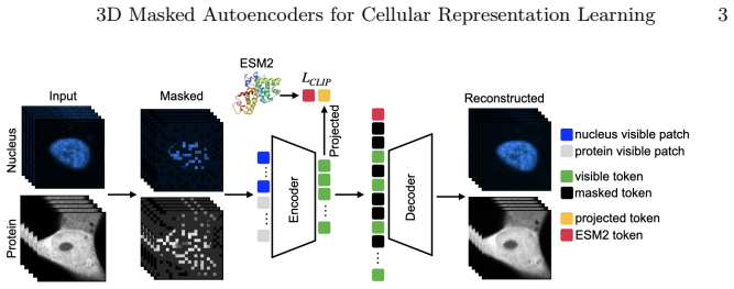
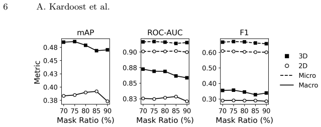
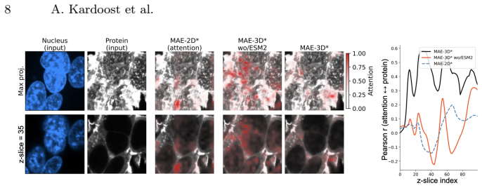
Under matched architectures and training protocols, MAE-3D consistently outperforms 2D max-projection and slice-based variants on downstream single-cell tasks. Cross-modal supervision with a pretrained protein language model yields larger gains for volumetric models. Channel cross-attention and frequency-domain regularization are critical for leveraging 3D spatial context. On a protein-protein interaction task, MAE-3D achieves a ROC-AUC of 0.865, outperforming prior methods by up to 0.025. For protein localization, the best 3D model attains state-of-the-art AUC_micro of 0.952 and F1_micro of 0.742, improving over previous approaches by 0.003 and 0.010 absolute, respectively.
What carries the argument
3D masked autoencoders with channel cross-attention and frequency-domain regularization, aligned to a pretrained protein language model through cross-modal supervision
Load-bearing premise
The observed performance gains arise from native 3D spatial context and cross-modal alignment rather than unstated differences in training protocols, data preprocessing, or model capacity.
What would settle it
Retraining the 2D and 3D models with identical data preprocessing, augmentation, hyperparameters, and compute budgets and finding no difference in downstream task performance.
Figures
read the original abstract
Self-supervised learning in fluorescence microscopy often relies on 2D projections, despite the inherently three-dimensional nature of cells. We present a systematic comparison of 2D and 3D masked autoencoders (MAE-2D vs. MAE-3D) on volumetric microscopy data. Under matched architectures and training protocols, MAE-3D consistently outperforms 2D max-projection and slice-based variants on downstream single-cell tasks. We further align visual representations with a pretrained protein language model (ESM2) and show that cross-modal supervision yields larger gains for volumetric models. Channel cross-attention and frequency-domain regularization are critical for leveraging 3D spatial context. On a protein--protein interaction task, MAE-3D achieves a ROC--AUC of 0.865, outperforming prior methods by up to +0.025. For protein localization, our best 3D model attains state-of-the-art AUC$_{\text{micro}}$ (0.952) and F1$_{\text{micro}}$ (0.742), improving over previous approaches by +0.003 and +0.010 absolute, respectively. Overall, these results demonstrate the advantages of native 3D modeling and multimodal alignment for representation learning in single-cell microscopy.
Editorial analysis
A structured set of objections, weighed in public.
Referee Report
Summary. The paper introduces 3D masked autoencoders (MAE-3D) for self-supervised representation learning on volumetric fluorescence microscopy data. It systematically compares them to 2D max-projection and slice-based MAE variants, claiming consistent outperformance under matched architectures and training protocols on downstream single-cell tasks. Cross-modal alignment with a pretrained ESM2 protein language model is shown to yield larger gains for 3D models, with channel cross-attention and frequency-domain regularization identified as key components. Reported results include a ROC-AUC of 0.865 on a protein-protein interaction task (+0.025 over priors) and state-of-the-art AUC_micro of 0.952 and F1_micro of 0.742 on protein localization (+0.003 and +0.010 absolute).
Significance. If the matched-conditions claim holds after verification, the work would establish native 3D modeling as advantageous for capturing volumetric spatial context in cellular microscopy representations, with multimodal alignment providing an additional benefit. This could influence self-supervised learning approaches in bioimaging by moving beyond 2D projections.
major comments (2)
- [Abstract] Abstract: The central claim that 'under matched architectures and training protocols, MAE-3D consistently outperforms' 2D variants is load-bearing for attributing gains to native 3D spatial context, yet the abstract asserts matching without enumerating concrete controls such as identical parameter counts, masking ratios, augmentations, optimizer schedules, or total FLOPs between variants. This detail is required to exclude hidden differences in capacity or data handling as the source of the +0.025 ROC-AUC and +0.010 F1_micro improvements.
- [Abstract] Abstract and implied Experiments section: The headline numerical results (ROC-AUC 0.865, AUC_micro 0.952, F1_micro 0.742) and claims of consistent outperformance are presented without error bars, standard deviations across runs, or statistical significance tests, preventing assessment of whether the reported gains are robust or could arise from experimental variability.
Simulated Author's Rebuttal
We thank the referee for the constructive feedback. We address each major comment below.
read point-by-point responses
-
Referee: [Abstract] Abstract: The central claim that 'under matched architectures and training protocols, MAE-3D consistently outperforms' 2D variants is load-bearing for attributing gains to native 3D spatial context, yet the abstract asserts matching without enumerating concrete controls such as identical parameter counts, masking ratios, augmentations, optimizer schedules, or total FLOPs between variants. This detail is required to exclude hidden differences in capacity or data handling as the source of the +0.025 ROC-AUC and +0.010 F1_micro improvements.
Authors: We agree that explicitly enumerating the matched controls would make the abstract more self-contained. The full manuscript details identical ViT-Base architectures (86M parameters), masking ratio 0.75, identical augmentations, AdamW optimizer with the same cosine schedule, and equivalent training epochs/FLOPs for MAE-2D and MAE-3D variants. We will revise the abstract to include a concise list of these controls. revision: yes
-
Referee: [Abstract] Abstract and implied Experiments section: The headline numerical results (ROC-AUC 0.865, AUC_micro 0.952, F1_micro 0.742) and claims of consistent outperformance are presented without error bars, standard deviations across runs, or statistical significance tests, preventing assessment of whether the reported gains are robust or could arise from experimental variability.
Authors: The abstract reports point estimates from primary runs. The experiments section reports means and standard deviations over five independent runs (different seeds) with paired t-test p-values for the gains. We will revise the abstract to report the metrics with standard deviations and note the statistical tests. revision: yes
Circularity Check
No significant circularity
full rationale
The paper is an empirical comparison of MAE-2D vs. MAE-3D on held-out downstream tasks (ROC-AUC 0.865, AUC_micro 0.952, F1_micro 0.742). No derivation chain, equations, or first-principles claims exist that reduce to fitted inputs or self-definitions. Performance metrics are standard external evaluations; the 'matched architectures' statement is a methodological assertion, not a circular reduction. No self-citation load-bearing steps or ansatz smuggling appear in the provided text.
Axiom & Free-Parameter Ledger
Reference graph
Works this paper leans on
-
[1]
In: 2024 IEEE/CVF Conference on Computer Vision and Pattern Recognition (CVPR)
Bourriez, N., Bendidi, I., Cohen, E., Watkinson, G., Sanchez, M., Bollot, G., Gen- ovesio, A.: Chada-vit: Channel adaptive attention for joint representation learning of heterogeneous microscopy images. In: Proceedings of the IEEE/CVF Conference on Computer Vision and Pattern Recognition (CVPR). pp. 11556–11565 (2024). https://doi.org/10.1109/CVPR52733.2024.01098
-
[2]
Bray, M.A., Singh, S., Han, H., Davis, C., Borgeson, B., Hartland, C., Kost- Alimova, M., Gustafsdottir, S.M., Gibson, C.C., Carpenter, A.E., et al.: The jump cell painting dataset: morphological impact of 136,000 chemical and genetic pertur- bations. Nature619, 151–158 (2023). https://doi.org/10.1038/s41586-023-06119-4
-
[3]
In: Proceedings of the 38th International Conference on Machine Learning (ICML)
Caron, M., Touvron, H., Misra, I., Jégou, H., Mairal, J., Bojanowski, P., Joulin, A.: Emerging properties in self-supervised vision transformers. In: Proceedings of the 38th International Conference on Machine Learning (ICML). pp. 2186–2205 (2021), http://proceedings.mlr.press/v139/caron21a.html
2021
-
[4]
In: ICML
Chen, T., Kornblith, S., Norouzi, M., Hinton, G.: A simple framework for con- trastive learning of visual representations. In: ICML. pp. 1597–1607 (2020)
2020
-
[5]
https://doi.org/10.1038/s41586-021-03969-3 10 A
Cho, N.H., Cheveralls, K.C., Brunner, A.D., Kim, K., Michaelis, A.C., Raghavan, P., Kobayashi, H., Savy, L., Li, J.Y., Canaj, H., et al.: Opencell: proteome-scale endogenoustaggingenablesthecartographyofhumancellularorganization.Nature 595, 285–290 (2021). https://doi.org/10.1038/s41586-021-03969-3 10 A. Kardoost et al
-
[6]
Doron, M., Moutakanni, T., Chen, Z.S., Moshkov, N., Caron, M., Touvron, H., Pernice, W., Caicedo, J.C.: Unbiased single-cell morphology with self-supervised vision transformers. bioRxiv (2023). https://doi.org/10.1101/2023.06.16.545359, https://www.biorxiv.org/content/10.1101/2023.06.16.545359v1
-
[7]
Dosovitskiy, A., Beyer, L., Kolesnikov, A., Weissenborn, D., Zhai, X., Unterthiner, T., Dehghani, M., Minderer, M., Heigold, G., Gelly, S., Uszkoreit, J., Houlsby, N.: An image is worth 16x16 words: Transformers for image recognition at scale. In: International Conference on Learning Representations (ICLR) (2021), https: //arxiv.org/abs/2010.11929
Pith/arXiv arXiv 2021
-
[8]
Gupta, A., Wefers, Z., Kahnert, K., Hansen, J.N., Misra, M.K., Leineweber, W., Cesnik, A., Lu, D., Axelsson, U., Ballllosera Navarro, F., Altman, R.B., Karaletsos, T., Lundberg, E.: Subcell: Vision foundation models for microscopy capture single- cell biology. bioRxiv (2024). https://doi.org/10.1101/2024.12.06.627299, https:// www.biorxiv.org/content/10.1...
-
[9]
He, K., Chen, X., Xie, S., Li, Y., Dollár, P., Girshick, R.: Masked autoencoders are scalablevisionlearners.In:ProceedingsoftheIEEE/CVFConferenceonComputer Vision and Pattern Recognition (CVPR). pp. 16000–16009 (2022). https://doi.org/ 10.1109/CVPR52688.2022.01553
-
[10]
In: ICCV (2021)
Jiang, L., Dai, B., Wu, W., Loy, C.C.: Focal frequency loss for image reconstruction and synthesis. In: ICCV (2021)
2021
-
[11]
Nature Methods19(8), 995–1003 (2022)
Kobayashi, H., Cheveralls, K.C., Leonetti, M.D., Royer, L.A., et al.: Self- supervised deep learning encodes high-resolution features of protein subcellular localization. Nature Methods19(8), 995–1003 (2022). https://doi.org/10.1038/ s41592-022-01541-z
2022
-
[12]
In: Proceedings of the IEEE/CVF Conference on Computer Vision and Pattern Recognition (CVPR)
Kraus, O., Kenyon-Dean, K., Saberian, S., Fallah, M., McLean, P., Leung, J., Sharma, V., Khan, A., Balakrishnan, J., Celik, S., Beaini, D., Sypetkowski, M., Cheng, C.V., Morse, K., Makes, M., Mabey, B., Earnshaw, B.: Masked autoen- coders for microscopy are scalable learners of cellular biology. In: Proceedings of the IEEE/CVF Conference on Computer Visio...
-
[13]
Evolutionary-scale prediction of atomic-level protein structure with a language model , volume =
Lin, Z., Akin, H., Rao, R., Hie, B., Zhu, Z., Lu, W., Smetanin, N., Verkuil, R., Kabeli, O., Shmueli, Y., dos Santos Costa, A., Fazel-Zarandi, M., Sercu, T., Candido, S., Rives, A.: Evolutionary-scale prediction of atomic-level protein structure with a language model. Science379(6637), 1123–1130 (2023). https: //doi.org/10.1126/science.ade2574, includes E...
-
[14]
PLOS Computational Biology21(12), e1013828 (2025)
Moutakanni, T., Couprie, C., Yi, S., Doron, M., Chen, Z.S., Moshkov, N., et al.: Cell-dino: Self-supervised image-based embeddings for cell fluorescent microscopy. PLOS Computational Biology21(12), e1013828 (2025). https://doi.org/10.1371/ journal.pcbi.1013828
2025
-
[15]
van den Oord, A., Vinyals, O., Kavukcuoglu, K.: Neural discrete representation learning.In:AdvancesinNeuralInformationProcessingSystems(NeurIPS).vol.30 (2017)
2017
-
[16]
Cellpose:ageneralistalgorithmforcellularsegmentation
Stringer, C., Wang, T., Michaelos, M., Pachitariu, M.: Cellpose: a generalist al- gorithm for cellular segmentation. Nature Methods18(1), 100–106 (2021). https: //doi.org/10.1038/s41592-020-01018-x
-
[17]
Science347(6220), 1260419 (2015)
Uhlén, M., Fagerberg, L., Hallström, B.M., Lindskog, C., Oksvold, P., Mardinoglu, A., Sivertsson, Å., Kampf, C., Sjöstedt, E., Asplund, A., et al.: Tissue-based map of the human proteome. Science347(6220), 1260419 (2015). https://doi.org/10. 1126/science.1260419 3D Masked Autoencoders for Cellular Representation Learning 11
2015
-
[18]
UniProt Consortium: Uniprot: the universal protein knowledgebase in 2023. Nu- cleic Acids Research51(D1), D523–D531 (2023). https://doi.org/10.1093/nar/ gkac1052
-
[19]
In: Advances in Neural Information Processing Systems (NeurIPS)
Vaswani,A.,Shazeer,N.,Parmar,N.,Uszkoreit,J.,Jones,L.,Gomez,A.N.,Kaiser, Ł., Polosukhin, I.: Attention is all you need. In: Advances in Neural Information Processing Systems (NeurIPS). vol. 30 (2017)
2017
-
[20]
Nature613(7943), 345–354 (2023)
Viana,M.P.,Chen,J.,Knijnenburg,T.A.,Vasan,R.,Yan,C.,Arakaki,J.E.,Bailey, M., Berry, B., Borensztejn, A., Brown, E.M., et al.: Integrated intracellular orga- nization and its variations in human ips cells. Nature613(7943), 345–354 (2023). https://doi.org/10.1038/s41586-022-05563-7
-
[21]
Nature Methods22(11), 2386–2399 (2025)
Zhou, F.Y., Marin, Z., Yapp, C., Zhou, Q., Nanes, B.A., Daetwyler, S., Jamieson, A.R., Islam, M.T., Jenkins, E., Gihana, G.M., Lin, J., Borges, H.M., Chang, B.J., Weems, A., Morrison, S.J., Sorger, P.K., Fiolka, R., Dean, K.M.: Universal consen- sus 3d segmentation of cells from 2d segmented stacks. Nature Methods22(11), 2386–2399 (2025). https://doi.org/...
-
[22]
Full wave inversion for ultrasound tomography using physics based deep neural network
Zhou, L., Liu, H., Bae, J., He, J., Samaras, D., Prasanna, P.: Self pre-training with masked autoencoders for medical image classification and segmentation. In: Pro- ceedings of the 2023 IEEE 20th International Symposium on Biomedical Imaging (ISBI). pp. 1–5 (2023). https://doi.org/10.1109/ISBI53787.2023.10230477, https: //ieeexplore.ieee.org/document/10230477
discussion (0)
Sign in with ORCID, Apple, or X to comment. Anyone can read and Pith papers without signing in.