REVIEW 2 major objections 6 minor 12 references
Rotational ultrasound sweeps lift prostate cancer AI past expert readers
Reviewed by Pith at T0; open to challenge. T0 means a machine referee read the full paper against a public rubric. the ladder, T0–T4 →
T0 review · glm-5.2
2026-07-09 22:54 UTC pith:LW3FURWG
load-bearing objection Compass fuses rotational sweep video with biopsy frames via a roll-angle-conditioned transformer for prostate cancer detection — the architecture is the real contribution, but the headline performance gap lacks significance testing. the 2 major comments →
Compass: Prostate Cancer Detection Needs Multi-View Context
The pith
A machine-rendered reading of the paper's core claim, the machinery that carries it, and where it could break.
Core claim
The core discovery is that fusing two distinct evidence streams—dense rotational prostate sweep frames and sparse biopsy-moment frames—through a single transformer that is conditioned on probe rotation angle produces substantially better patient-level cancer detection than either stream alone or any single-frame, MIL, or video baseline tested. The mechanism is cross-branch attention: sweep tokens and biopsy tokens attend to each other within one shared sequence, allowing localized biopsy evidence to align with rotation-consistent sweep patterns.
What carries the argument
The architecture has four moving parts. First, a frozen image encoder (ProstNFound+) produces visual tokens for each sweep frame and each biopsy frame. Second, a sinusoidal positional encoding embeds the probe's continuous roll angle into each token, giving the model geometric awareness of which rotational view it is processing. Third, two lightweight decoders—one for sweep frames, one for biopsy frames—compress their respective token sets and add the roll embedding plus branch-specific shared tokens. Fourth, all tokens from both branches are concatenated with a learnable classification token and passed through a transformer encoder whose global self-attention lets every token attend to all.
Load-bearing premise
The central performance claim rests on the assumption that a 118-patient, two-center cohort is large enough to generalize the reported 87.2% AUROC and the 8.7-point improvement over the next-best baseline, even though the standard deviations are large and no statistical significance tests are reported.
What would settle it
If, on a larger independent cohort, Compass's patient-level AUROC drops to within the confidence interval of single-frame baselines or expert PRI-MUS readers, the claim that multi-view fusion provides substantial added value would be falsified. Likewise, if the cross-branch transformer ablation gap vanishes with more data, the specific mechanism of cross-branch reasoning would be undermined.
If this is right
- If multi-view sweep fusion genuinely improves patient-level triage, clinics could use Compass as a pre-biopsy risk stratification tool, potentially reducing unnecessary biopsies for low-risk patients.
- The cross-branch transformer design—pairing dense contextual data with sparse confirmed samples—could transfer to other medical imaging tasks where a global scan coexists with targeted tissue sampling, such as breast ultrasound or liver imaging.
- Roll-angle conditioning as a positional signal could generalize to any probe-based imaging modality where sensor orientation is available, enabling rotation-aware reasoning in endoscopic or intravascular ultrasound.
Where Pith is reading between the lines
- The paper does not report statistical significance tests for the AUROC gap between Compass and the next-best baseline; given the small cohort (118 patients) and large standard deviations, the headline improvement may not be statistically robust. A reader should treat the 8.7-point AUROC gap as suggestive rather than proven.
- The claim that cross-branch reasoning specifically drives the gain is supported by the ablation, but the ablation itself is also subject to the same small-sample variance, so the causal attribution to the transformer mechanism is not fully decoupled from sample-size noise.
- If the approach scales to larger cohorts and the improvement holds, it would shift the design paradigm for ultrasound-based cancer detection AI from frame-level classifiers toward study-level fusion architectures, which has architectural and computational implications for real-time deployment.
Editorial analysis
A structured set of objections, weighed in public.
Referee Report
Summary. The paper proposes Compass, a multi-view framework for prostate cancer (PCa) detection from micro-ultrasound (μUS). Compass integrates two evidence streams—rotational sweep cine-loops and biopsy-core frames—using a transformer encoder conditioned on the probe's roll angle, producing both patient-level and biopsy-level risk scores. The method is evaluated on a 118-patient, 2-center subset of the OPTIMUM clinical trial, and compared against frame-level classifiers, MIL variants, video models, and expert PRI-MUS readers. Compass achieves 87.2% patient-level AUROC, outperforming the best baseline (ProstNFound+ at 78.7%) and expert readers (78.5%). The architecture is well-motivated by clinical workflow, and ablation results support the contribution of each component.
Significance. The paper addresses a genuine gap in AI-based μUS PCa detection: most prior methods analyze single frames in isolation, while clinicians interpret full 3D sweep studies. The dual-branch design—fusing sweep context with biopsy frames via roll-angle-conditioned cross-attention—is a reasonable and novel architectural contribution. The use of a multi-center clinical trial dataset (OPTIMUM) and comparison against expert PRI-MUS scores adds clinical credibility. The authors provide publicly available code, which is a notable strength for reproducibility. The ablation study (Table 2) provides internally consistent evidence that sweeps, angle encoding, and the transformer each contribute to performance.
major comments (2)
- §5, Table 1: No paired statistical significance tests are reported for the headline AUROC comparisons. Compass (87.2±8.7) vs. ProstNFound+ (78.7±15.4) is the comparison most directly supporting the central claim of 'outperforming' baselines. Because both methods are evaluated on identical 5-fold cross-validation splits, a paired test (e.g., paired bootstrap of per-fold AUROC differences, or DeLong test per fold) would be the appropriate and more powerful check. The reported standard deviations are large and plausibly overlapping; without a paired comparison, the 8.5-point gap cannot be distinguished from small-sample noise. The same concern applies to the ablation results in Table 2 (e.g., Compass 87.2 vs. 'No sweeps' 78.8), though the effect sizes there are larger and more internally convincing. The authors should report per-fold paired significance tests for at least the primary and ab
- §3, 'Sweep-level feature extraction': The backbone encoder ProstNFound+ [11] is prior work by overlapping author groups. The paper states it was chosen 'due to its strong performance on and robust prostate US representations' but does not disclose whether ProstNFound+ was trained or fine-tuned on data from the OPTIMUM trial or any data overlapping with the 118-patient cohort used here. If overlap exists, it should be disclosed. Note that this concern is mitigated for the Compass-vs-ProstNFound+ comparison (both use the same frozen backbone on the same data), but it matters for absolute performance claims and for the comparison against expert readers. The authors should explicitly state the training data provenance of the backbone.
minor comments (6)
- §4, 'Baselines & Evaluation': The criteria for selecting the 'epoch with the best mean patient AUROC across folds' for biopsy-level metrics could introduce optimistic bias if the epoch is selected using the same metric being reported. Clarify whether epoch selection used a held-out criterion.
- Table 1: The standard deviations for some entries are very large (e.g., ProstNFound+ ±15.4 AUROC, MedSAM Sen@60 ±24.4). Reporting per-fold results or confidence intervals would help readers assess stability.
- Figure 1: The diagram is dense and some labels are difficult to parse at standard resolution. Consider enlarging or simplifying the annotation of the cross-attention and self-attention modules.
- §3, 'Loss function': The value of λ=0.05 is stated in §4 but not motivated. A brief justification or sensitivity analysis would strengthen the claim that this hyperparameter is not finely tuned.
- §2: The dataset description mentions 'at least one initial transverse scan' per study. It is unclear how many sweep frames T are used per patient on average, and whether T is fixed or variable. This affects interpretability of the transformer input length.
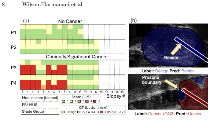
- Figure 3a: The axis labels and legend are small. The meaning of 'P1–P4' and the row groupings could be stated more explicitly in the caption.
Circularity Check
No circularity found; the model's predictions are not defined by its inputs by construction.
full rationale
The paper proposes Compass, a transformer-based architecture for prostate cancer detection. The derivation chain is straightforward: a frozen image encoder (ProstNFound+) produces visual tokens from sweep and biopsy frames; these tokens are combined with roll-angle positional encodings and clinical metadata prompts; a transformer encoder performs self-attention over the mixed-token sequence; and decoder heads predict patient-level and biopsy-level risk scores. The loss function combines patient-level cross-entropy with biopsy-level cross-entropy. There is no step where the prediction is defined in terms of the label, and no fitted parameter is renamed as a prediction. The reader's concern about potential data overlap with the ProstNFound+ backbone is a validity/generalization concern (correctness risk), not a circularity in the derivation chain. The ablation study (Table 2) shows that removing components degrades performance, confirming the outputs are not trivially forced by the inputs. The self-citation to ProstNFound+ [11] is used to provide a feature extractor, not to invoke a uniqueness theorem or smuggle an ansatz. The paper is self-contained against external benchmarks (PRI-MUS experts, CLIP, MedSAM). No circularity is present.
Axiom & Free-Parameter Ledger
free parameters (5)
- λ (biopsy loss weight) =
0.05
- Learning rate =
3e-5
- d (token dimension) =
256
- τ_sw, τ_bx (shared token embeddings)
- [CLS] token initialization
axioms (4)
- domain assumption ProstNFound+ produces robust prostate US representations suitable as frozen backbone features.
- domain assumption The roll angle from the inertial measurement unit provides a reliable approximation of the left-right position of the imaging plane.
- domain assumption The frame immediately preceding needle firing is a canonical representation of the biopsy core's tissue context.
- domain assumption 5-fold patient-level cross-validation on 118 patients provides reliable performance estimates for model comparison.
read the original abstract
Artificial intelligence (AI) analysis of micro-ultrasound ($\mu$US) has shown promise for prostate cancer (PCa) detection. However, most existing AI methods focus on the analysis of single $\mu$US images in isolation. By contrast, expert $\mu$US readers typically assess a full recorded video study, which provides three-dimensional context, to improve PCa detection compared to single-frame analysis. Inspired by this clinical workflow, we propose Compass, a novel AI methodology which models a $\mu$US study as a stream of 2D images. Compass jointly integrates rotational sweep videos of the prostate with $\mu$US frames acquired at the moment of biopsy, and performs evidence aggregation across the study using a transformer conditioned on the probe's rotational angle. Finally, a decoder head predicts frame-level and study-level risk scores for the patient. The model is trained and evaluated using a multi-center clinical trial dataset of $\mu$US studies, including continuous rotational scans of the prostate and videos captured during biopsy acquisition. We compare the proposed method to baseline AI methods from the literature and to risk scores provided by clinical experts. Our framework shows strong performance, highlighting the value of multi-view context for $\mu$US PCa detection, and providing a potentially powerful tool to complement human expertise in $\mu$US-based PCa diagnosis. Our code is available at: https://github.com/mharmanani/Compass.
Figures


Reference graph
Works this paper leans on
-
[1]
In: Proceedings of the IEEE/CVF international confer- ence on computer vision
Arnab, A., Dehghani, M., Heigold, G., Sun, C., Lučić, M., Schmid, C.: Vivit: A video vision transformer. In: Proceedings of the IEEE/CVF international confer- ence on computer vision. pp. 6836–6846 (2021)
work page 2021
-
[2]
In: International Conference on Medical Image Computing and Computer-Assisted Intervention
Elghareb, T., Harmanani, M., To, M.N.N., Wilson, P., Jamzad, A., Fooladgar, F., Abdelsamad, B., Dzikunu, O., Sojoudi, S., Reznik, G., et al.: Proteus: A spatio- temporal enhanced ultrasound-based framework for prostate cancer detection. In: International Conference on Medical Image Computing and Computer-Assisted Intervention. pp. 405–414. Springer (2025)
work page 2025
-
[3]
The Journal of Urology196(2), 562–569 (Aug 2016)
Ghai, S., Eure, G., Fradet, V., Hyndman, M.E., McGrath, T., Wodlinger, B., Pavlovich, C.P.: Assessing Cancer Risk on Novel 29 MHz Micro-Ultrasound Im- ages of the Prostate: Creation of the Micro-Ultrasound Protocol for Prostate Risk Identification. The Journal of Urology196(2), 562–569 (Aug 2016). https://doi.org/10.1016/j.juro.2015.12.093
-
[4]
In: 2025 IEEE 22nd International Symposium on Biomedical Imaging (ISBI)
Harmanani, M., Jamzad, A., To, M.N.N., Wilson, P.F., Guo, Z., Fooladgar, F., Sojoudi, S., Gilany, M., Chang, S., Black, P., et al.: Cinepro: Robust training of foundation models for cancer detection in prostate ultrasound cineloops. In: 2025 IEEE 22nd International Symposium on Biomedical Imaging (ISBI). pp. 1–5. IEEE (2025)
work page 2025
-
[5]
BJUI Compass7(2), e70133 (2026)
Imran, M., Brisbane, W.G., Su, L.M., Joseph, J.P., Shao, W.: Ai-enhanced micro- ultrasound improves detection of clinically significant prostate cancer at biopsy. BJUI Compass7(2), e70133 (2026)
work page 2026
-
[6]
Computerized Medical Imaging and Graphics112, 102326 (2024)
Jiang, H., Imran, M., Muralidharan, P., Patel, A., Pensa, J., Liang, M., Beni- dir, T., Grajo, J.R., Joseph, J.P., Terry, R., et al.: Microsegnet: A deep learning approach for prostate segmentation on micro-ultrasound images. Computerized Medical Imaging and Graphics112, 102326 (2024)
work page 2024
-
[7]
JAMA333(19), 1679–1687 (May 2025)
Kinnaird, A., Luger, F., Cash, H., Ghai, S., Urdaneta-Salegui, L.F., Pavlovich, C.P., Brito, J., Shore, N.D., Struck, J.P., Schostak, M., others, OPTIMUM Inves- tigators:Microultrasonography-GuidedvsMRI-GuidedBiopsyforProstateCancer Diagnosis: The OPTIMUM Randomized Clinical Trial. JAMA333(19), 1679–1687 (May 2025). https://doi.org/10.1001/jama.2025.3579 ...
-
[8]
Lee, J.H., Li, C.X., Jahanandish, H., Bhattacharya, I., Vesal, S., Zhang, L., Sang, S., Choi, M.H., Soerensen, S.J.C., Zhou, S.R., Sommer, E.R., Fan, R., Ghanouni, P., Song, Y., Seibert, T.M., Sonn, G.A., Rusu, M.: Prostate-specific foundation models for enhanced detection of clinically significant cancer (2025), https://arxiv.org/abs/2502.00366
work page internal anchor Pith review Pith/arXiv arXiv 2025
-
[9]
European Urology87(3), 302–313 (2025)
Schafer, E.J., Laversanne, M., Sung, H., Soerjomataram, I., Briganti, A., Dahut, W., Bray, F., Jemal, A.: Recent patterns and trends in global prostate cancer incidence and mortality: an update. European Urology87(3), 302–313 (2025)
work page 2025
-
[10]
Nature Cancer 6(10), 1621–1637 (Oct 2025)
Shao, L., Liang, C., Yan, Y., Zhu, H., Jiang, X., Bao, M., Zang, P., Huang, X., Zhou,H.,Nie,P.,Wang,L.,Li,J.,Zhang,S.,Ren,S.:Anmri–pathologyfoundation model for noninvasive diagnosis and grading of prostate cancer. Nature Cancer 6(10), 1621–1637 (Oct 2025). https://doi.org/10.1038/s43018-025-01041-x, epub 2025-09-02
-
[11]
International Journal of Computer Assisted Radiology and Surgery (Feb 2026)
Wilson, P.F.R., Harmanani, M., To, M.N.N., Jamzad, A., Elghareb, T., Guo, Z., Kinnaird, A., Wodlinger, B., Abolmaesumi, P., Mousavi, P.: ProstNFound+: A prospective study using medical foundation models for prostate cancer detection. International Journal of Computer Assisted Radiology and Surgery (Feb 2026). https://doi.org/10.1007/s11548-025-03561-4
-
[12]
In: International conference on medical image computing and computer-assisted intervention
Wilson, P.F., To, M.N.N., Jamzad, A., Gilany, M., Harmanani, M., Elghareb, T., Fooladgar, F., Wodlinger, B., Abolmaesumi, P., Mousavi, P.: Prostnfound: inte- grating foundation models with ultrasound domain knowledge and clinical context for robust prostate cancer detection. In: International conference on medical image computing and computer-assisted int...
work page 2024
discussion (0)
Sign in with ORCID, Apple, or X to comment. Anyone can read and Pith papers without signing in.