REVIEW 2 major objections 5 minor 152 references
AI in nanoparticle electron microscopy is shifting from labeling what is visible to recovering structure, dynamics, and materials insight.
Reviewed by Pith at T0; open to challenge. T0 means a machine referee read the full paper against a public rubric. the ladder, T0–T4 →
T0 review · grok-4.5
2026-07-14 12:05 UTC pith:N35EQ5CQ
load-bearing objection Solid challenge-organized review of AI for nanoparticle EM; useful taxonomy and fidelity caveats, ordinary review-selection risk on the tables. the 2 major comments →
The evolution of AI from image interpretation toward scientific inference in nanoparticle electron microscopy
The pith
A machine-rendered reading of the paper's core claim, the machinery that carries it, and where it could break.
Core claim
AI methodologies in nanoparticle electron microscopy have evolved from image-level interpretation—detection and segmentation—into tools for scientific inference: recovering lattice-level structure under noise, estimating latent three-dimensional morphology from projections, and quantifying dynamic nanoscale processes, by coupling computer vision with microscopy physics, simulation-based training, metadata, and increasingly autonomous experimental control.
What carries the argument
A hierarchical task ladder from detection and segmentation, through atomic-resolution restoration, to 2D-to-3D structural inference and spatiotemporal analysis of in situ data, with physics-informed and simulation-driven learning as the bridge from visible pixels to latent physical variables.
Load-bearing premise
That the reported performance ranges and maturity levels across detection, denoising, structural inference, and dynamics fairly represent the broader literature, even though many high scores rest on simulated ground truth or carefully curated datasets that may not match routine experimental conditions.
What would settle it
A community benchmark that holds the same models to experimental nanoparticle TEM, HRTEM, and in situ video sets with independent physical validation (for example measured growth rates, facet statistics, or strain maps) and shows that the claimed progression from image labeling to reliable scientific inference does not hold outside simulation or narrow curated tests.
If this is right
- Detection and segmentation become routine infrastructure, while the competitive frontier shifts to restoration fidelity, uncertainty-aware 3D inference, and kinetic quantification from video.
- Physics-informed and hybrid simulation–experiment training becomes standard for atomic-resolution and inverse problems because pure data-driven models lack experimental ground truth.
- Benchmarking moves from PSNR, Dice, and mAP toward structural fidelity metrics (such as Fourier ring correlation) and measurable physical outputs (diffusion coefficients, coalescence rates, facet distributions).
- Foundation models, multimodal image–metadata systems, and closed-loop AI control turn microscopes into active discovery platforms rather than post-processing cameras.
- Materials discovery pipelines can treat AI-extracted nanoparticle descriptors as direct inputs to structure–property models and inverse design.
Where Pith is reading between the lines
- If structural inference and dynamics remain under-benchmarked relative to detection, the field risks overstating readiness for autonomous materials discovery while the load-bearing inverse problems stay unstandardized.
- The same hierarchical ladder implies that progress on rare-event kinetics and defect energetics will depend more on uncertainty quantification and domain adaptation than on larger generic vision backbones alone.
- Commercial AI microscopy platforms will face pressure to publish transparent experimental benchmarks, not only internal Dice or mAP numbers, if the review’s maturity map is taken seriously.
Editorial analysis
A structured set of objections, weighed in public.
Referee Report
Summary. This review argues that AI for nanoparticle electron microscopy has progressed from image-level interpretation (detection/segmentation of TEM ensembles) toward scientific inference: atomic-resolution restoration with structural fidelity, 2D-to-3D structural inference, spatiotemporal analysis of in situ dynamics, and closed-loop autonomous experimentation. The narrative is organized by scientific challenge rather than by algorithm family (Fig. 2; §§4–7), covering TEM/HRTEM/STEM and in situ TEM, and spanning CNNs through transformers, self-supervised learning, foundation models, multimodal AI, and physics-informed/simulation-driven methods. Representative performance and evaluation tables (Tables 1–4) and architecture appendices (Tables A1–A4) support maturity rankings and a forward-looking agenda (Table 5; §8).
Significance. If the synthesis is accepted, the paper provides a useful, problem-centered map of a rapidly expanding and fragmented literature for materials scientists and microscopists. Strengths include the hierarchical framing (Fig. 2), repeated emphasis on simulation-to-experiment domain gap, structural fidelity over PSNR/SSIM (FRC, crystallographic consistency), and the need for uncertainty quantification and community benchmarks (§§5.4, 6.3, 7.4, 8). The appendices (A1–A4) and commercial-tool mention give practical orientation. As a review, its value is organizational and critical rather than a new theorem or experiment; residual risk is ordinary selection/maturity bias in aggregated metrics, which the manuscript already flags.
major comments (2)
- Table 1 and §4.5 present typical mAP/Dice/IoU ranges (e.g., Dice 0.85–0.95, mAP >0.90) as evidence of maturity. The text correctly notes dependence on morphology, contrast, and annotation, but the table still reads as cross-study comparable. Please state explicitly that ranges are illustrative upper bounds from curated datasets (often with simulated or expert labels), not meta-analytic estimates, and add a short note on how many studies and what domain shifts underlie each row so maturity rankings cannot be over-read.
- Tables 2–4 and §§5.4, 6.3, 7.4 correctly stress missing experimental ground truth and domain shift, yet Table 5 and §8 still rank detection/segmentation as “highly mature” and list foundation models/closed-loop systems as primary next drivers. Tighten the link: either qualify maturity labels with “on curated/static TEM” vs “atomic/dynamic/inverse tasks,” or add one paragraph that separates (i) routine morphology pipelines from (ii) inference tasks where simulation fidelity and uncertainty remain load-bearing, so the evolutionary claim is not overstated by the most mature subfield alone.
minor comments (5)
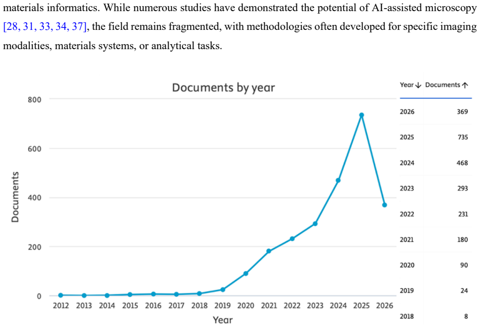
- Figure 1 caption and text cite Scopus analysis “performed in April 2026” and a 2025 peak of 735 papers; ensure the query string, date, and counts are reproducible and consistent with the arXiv posting timeline.
- Several figure panels (Figs. 3–7, A1) are author-generated schematics “inspired by” literature; captions should state more clearly that they are illustrative, not reprocessed experimental data, to avoid misreading as primary results.
- Reference list has occasional incomplete or inconsistent entries (e.g., missing DOIs or venue details for some 2025–2026 items; Table A4 “Giner et al.” with “—” DOI). Normalize citations and fix typographical issues (e.g., “Heslignton,” duplicated sensing refs [7, 12, 12]).
- Appendix Tables A1–A3 are valuable but dense; a brief caveat that “First Use in Microscopy” is a class of applications rather than a single definitive paper (already noted in text) should appear in the table caption for skimmers.
- Commercial platform SenseAI is listed in Table A4/Table 5; a one-sentence disclosure of any author relationship (or none) would improve transparency for readers.
Circularity Check
No circular derivation: this is a hierarchical literature review whose evolutionary narrative is synthesis of external work, not a self-referential prediction or fitted result.
full rationale
The manuscript is a review organized around a hierarchy of AI tasks in nanoparticle electron microscopy (detection/segmentation → atomic restoration → 2D-to-3D inference → spatiotemporal dynamics → scientific inference; Abstract; §§1–3, 8; Fig. 2). It does not claim a first-principles derivation, uniqueness theorem, or parameter-free prediction that could reduce to its own inputs. Representative performance ranges (Tables 1–4) and architecture timelines (Tables A1–A4) are presented as literature summaries with explicit caveats on simulated ground truth, domain shift, missing community benchmarks, and hallucination risk (§§4.5, 5.4, 6.3, 7.4). Author-related citations (e.g., nanoparticle synthesis/microscopy background) are ordinary context and do not force the review’s conclusions by construction. No self-definitional equation, fitted-input-called-prediction, load-bearing self-citation uniqueness claim, or renamed known result appears. Circularity score is therefore 0.
Axiom & Free-Parameter Ledger
axioms (4)
- domain assumption TEM/STEM images are 2D projections of 3D nanostructures whose contrast is governed by electron scattering, dose, aberrations, thickness, and orientation.
- domain assumption Experimentally verified noise-free atomic-resolution ground truth is generally unavailable, so simulation-driven or self-supervised training is necessary.
- ad hoc to paper Reported detection/segmentation metrics (mAP, Dice, IoU) and restoration metrics (PSNR, SSIM, FRC) are sufficiently comparable across studies to support maturity rankings.
- ad hoc to paper Foundation models, multimodal learning, and closed-loop autonomous microscopy will become primary drivers of next-generation nanoparticle characterization.
read the original abstract
Artificial intelligence (AI) is transforming electron microscopy by enabling quantitative analysis of increasingly large and complex datasets for nanoparticle characterization. Recent advances in machine learning (ML) and deep learning (DL) have expanded microscopy from a descriptive imaging technique into a data-driven platform for structural interpretation, dynamic analysis, and scientific inference. This review examines AI methodologies for nanoparticle electron microscopy, focusing on transmission electron microscopy (TEM), high-resolution transmission electron microscopy (HRTEM), scanning transmission electron microscopy (STEM), and in situ TEM. The discussion is organized around the principal challenges in nanoparticle characterization, including particle detection, segmentation, morphology quantification, atomic-resolution restoration, defect identification, two-dimensional-to-three-dimensional structural inference, and analysis of dynamic processes in situ. We review computational approaches from conventional ML and convolutional neural networks to transformer architectures, self-supervised learning, foundation models, multimodal AI, and physics-informed learning. We further discuss integrating microscopy data with simulations, metadata, and autonomous experimentation to relate nanoparticle structure, dynamics, synthesis conditions, and functional properties. The advantages, limitations, benchmarking, and data requirements of current methodologies are critically assessed. Finally, emerging opportunities for foundation models, AI-guided microscopy, closed-loop experimentation, and autonomous materials discovery are discussed. By integrating advances across computer vision, materials informatics, and electron microscopy, this review highlights the role of AI in next-generation nanoparticle characterization and accelerated materials discovery.
Figures


Reference graph
Works this paper leans on
-
[1]
Nanoparticle Design by Gas-Phase Synthesis
Grammatikopoulos, P.; Steinhauer, S.; Vernieres, J.; Singh, V.; Sowwan, M. Nanoparticle Design by Gas-Phase Synthesis. Adv. Phys. X 2016, 1, 81–100. https://doi.org/10.1080/23746149.2016.1142829
-
[2]
Metal Catalysts for Heterogeneous Catalysis: From Single Atoms to Nanoclusters and Nanoparticles
Liu, L.; Corma, A. Metal Catalysts for Heterogeneous Catalysis: From Single Atoms to Nanoclusters and Nanoparticles. Chem. Rev. 2018, 118, 4981–5079. https://doi.org/10.1021/acs.chemrev.7b00776
-
[3]
Farooq, N.; Rehman, Z. U.; Khan, M. I.; et al. Nanomaterial-Based Energy Conversion and Energy Storage Devices: A Comprehensive Review. New J. Chem. 2024, 48, 8933. https://doi.org/10.1039/D3NJ04846B
-
[4]
Anker, J. N.; Hall, W. P.; Lyandres, O.; Shah, N. C.; Zhao, J.; Van Duyne, R. P. Biosensing with Plasmonic Nanosensors. Nat. Mater. 2008, 7, 442–453. https://doi.org/10.1038/nmat2162
doi:10.1038/nmat2162 2008
-
[5]
Magnetic Nanoparticles: Synthesis, Anisotropy, and Applications
Ma, Z.; et al. Magnetic Nanoparticles: Synthesis, Anisotropy, and Applications. Chem. Rev. 2022, 122, 127–231. https://doi.org/10.1021/acs.chemrev.1c00860
-
[6]
McNamara, K.; Tofail, S. A. M. Nanoparticles in Biomedical Applications. Adv. Phys. X 2017, 2, 54–
2017
-
[7]
https://doi.org/10.1080/23746149.2016.1254570
-
[8]
Mayer, K. M.; Hafner, J. H. Localized Surface Plasmon Resonance Sensors. Chem. Rev. 2011, 111, 3828–3857. https://doi.org/10.1021/cr100313v
-
[9]
Bhattacharya, S.; Gupta, A. B.; Gupta, A.; Pandey, A. A Review on Nanomaterials for Environmental Remediation. Energy Environ. Sci. 2012, 5, 8075–8109. https://doi.org/10.1039/C2EE21818F
-
[10]
L.; Seifer, S.; Ilavsky, J.; Pellin, M.; Sowwan, M.; Grammatikopoulos, P.; Vajda, S
Halder, A.; Kioseoglou, J.; Yang, B.; Kolipaka, K. L.; Seifer, S.; Ilavsky, J.; Pellin, M.; Sowwan, M.; Grammatikopoulos, P.; Vajda, S. Nanoassemblies of Ultrasmall Clusters with Remarkable Activity in 43 Carbon Dioxide Conversion to C1 Fuels. Nanoscale 2019, 11, 4683–4687. https://doi.org/10.1039/C8NR06664G
-
[11]
Hydrogen Flux through Size-Selected Pd Nanoparticles into Underlying Mg Nanofilms
Kumar, S.; Pavloudis, T.; Singh, V.; Nguyen, S.; Steinhauer, S.; Pursell, C.; Clemens, B.; Kioseoglou, J.; Grammatikopoulos, P.; Sowwan, M. Hydrogen Flux through Size-Selected Pd Nanoparticles into Underlying Mg Nanofilms. Adv. Energy Mater. 2018, 8, 1701326. https://doi.org/10.1002/aenm.201701326
-
[12]
S.; Mathioudaki, S.; Kioseoglou, J.; Grammatikopoulos, P
Bohra, M.; Giaremis, S.; Abisegapriyan, K. S.; Mathioudaki, S.; Kioseoglou, J.; Grammatikopoulos, P. Ferromagnetic-Antiferromagnetic Coupling in Gas-Phase Synthesized M(Fe, Co, Ni)-Cr Nanoparticles for Next-Generation Magnetic Applications. Adv. Sci. 2024, 11, 2403708. https://doi.org/10.1002/advs.202403708
-
[13]
Sosada-Ludwikowska, F.; Reiner, L.; Egger, L.; Lackner, E.; Krainer, J.; Wimmer-Teubenbacher, R.; Singh, V.; Steinhauer, S.; Grammatikopoulos, P.; Köck, A. Adjusting Surface Coverage of Pt Nanocatalyst Decoration for Selectivity Control in CMOS-Integrated SnO₂ Thin Film Sensors. Nanoscale Adv. 2024, 6, 1127–1134. https://doi.org/10.1039/D3NA00552F
-
[14]
E.; Nordlund, K.; Djurabekova, F.; Grammatikopoulos, P.; Sowwan, M
Vernieres, J.; Steinhauer, S.; Zhao, J.; Chapelle, A.; Menini, P.; Dufour, N.; Diaz, R. E.; Nordlund, K.; Djurabekova, F.; Grammatikopoulos, P.; Sowwan, M. Gas Phase Synthesis of Multifunctional Fe- Based Nanocubes. Adv. Funct. Mater. 2017, 27, 1605328. https://doi.org/10.1002/adfm.201605328
-
[15]
B.; Carter, C
Williams, D. B.; Carter, C. B. Transmission Electron Microscopy: A Textbook for Materials Science; Springer: New York, 2009
2009
-
[16]
J.; Nellist, P
Pennycook, S. J.; Nellist, P. D., Eds. Scanning Transmission Electron Microscopy: Imaging and Analysis; Springer: New York, 2011
2011
-
[17]
Genç, A.; Marlowe, J.; Jalil, A.; Belzberg, D.; Kovarik, L.; Christopher, P. A Versatile Machine Learning Workflow for High-Throughput Analysis of Supported Metal Catalyst Particles. Ultramicroscopy 2025, 271, 114116. https://doi.org/10.1016/j.ultramic.2025.114116
-
[18]
Lee, B.; et al. Statistical Characterization of the Morphologies of Nanoparticles through Machine Learning Based Electron Microscopy Image Analysis. ACS Nano 2020, 14, 17125–17133. https://doi.org/10.1021/acsnano.0c06809
-
[19]
Ziatdinov, M.; Dyck, O.; Jesse, S.; Kalinin, S. V. Deep Learning of Atomically Resolved Scanning Transmission Electron Microscopy Data. ACS Nano 2017, 11:12, 12742-12752. https://doi.org/10.1021/acsnano.7b07504
-
[20]
Madsen, J.; Liu, P.; Kling, J.; Wagner, J. B.; Hansen, T. W.; Winther, O.; Schiøtz, J. A. Deep Learning Approach to Identify Local Structures in Atomic-Resolution Transmission Electron Microscopy Images. Adv. Theory Simul. 2018, 1, 1800037. https://doi.org/10.1002/adts.201800037
-
[21]
Ophus, C. Four-Dimensional Scanning Transmission Electron Microscopy (4D-STEM): From Scanning Nanodiffraction to Ptychography and Beyond. Microsc. Microanal. 2019, 25, 563–582. https://doi.org/10.1017/S1431927619000497
-
[22]
Characterization of Nanomaterials Using In Situ Liquid-Cell Transmission Electron Microscopy
Chen, Y.; et al. Characterization of Nanomaterials Using In Situ Liquid-Cell Transmission Electron Microscopy. ACS Appl. Nano Mater. 2023, 6, 24, 22545–22567. https://doi.org/10.1021/acsanm.3c03003 44
-
[23]
Recent Advances in In Situ Transmission Electron Microscopy for Heterogeneous Catalysis
Qu, J.; et al. Recent Advances in In Situ Transmission Electron Microscopy for Heterogeneous Catalysis. Chem. Soc. Rev. 2023, 26, 7, 107072. https://doi.org/10.1016/j.isci.2023.107072
-
[24]
Martineau, B. H.; Johnstone, D. N.; van Helvoort, A. T. J.; et al. Unsupervised Machine Learning Applied to Scanning Precession Electron Diffraction Data. Adv. Struct. Chem. Imaging 2019, 5, 3. https://doi.org/10.1186/s40679-019-0063-3
-
[25]
Scanning Precession Electron Diffraction—Beyond Orientation and Phase Mapping
Bergh, T. Scanning Precession Electron Diffraction—Beyond Orientation and Phase Mapping. Microsc. Microanal. 2025, 31 (Suppl. 1), ozaf048.288. https://doi.org/10.1093/mam/ozaf048.288
-
[26]
Gai, P. L.; Lari, L.; Ward, M. R.; Boyes, E. D. Visualisation of Single Atom Dynamics and their Role in Nanocatalysts under Controlled Reaction Environments. Chem. Phys. Lett. 2014, 592, 355–359. https://doi.org/10.1016/j.cplett.2013.12.038
-
[27]
Steinhauer, S.; Vernieres, J.; Krainer, J.; Köck, A.; Grammatikopoulos, P.; Sowwan, M. In Situ Chemoresistive Sensing in the Environmental TEM: Probing Functional Devices and their Nanoscale Morphology. Nanoscale 2017, 9, 7380–7384. https://doi.org/10.1039/C6NR09322A
-
[28]
Steinhauer, S.; Zhao, J.; Singh, V.; Pavloudis, T.; Kioseoglou, J.; Nordlund, K.; Djurabekova, F.; Grammatikopoulos, P.; Sowwan, M. Thermal Oxidation of Size-Selected Pd Nanoparticles Supported on CuO Nanowires: The Role of the CuO–Pd Interface. Chem. Mater. 2017, 29, 6153–6160. https://doi.org/10.1021/acs.chemmater.7b02242
-
[29]
Botifoll, M.; et al. Machine Learning in Electron Microscopy for Advanced Nanocharacterization: Current Developments, Available Tools and Future Outlook. Nanoscale Horiz. 2022, 7, 1427–1477. https://doi.org/10.1039/D2NH00377E
-
[30]
Dynamic Wet-ETEM Observation of Pt/C Electrode Catalysts in a Moisturized Cathode Atmosphere
Yoshida, K.; et al. Dynamic Wet-ETEM Observation of Pt/C Electrode Catalysts in a Moisturized Cathode Atmosphere. Nanotechnology 2014, 25, 425702. https://doi.org/10.1088/0957- 4484/25/42/425702
doi:10.1088/0957- 2014
-
[31]
Dearg, M.; Roncaglia, C.; Nelli, D.; El Koraychy, E. Y.; Ferrando, R.; Slater, T. J. A.; Palmer, R. E. Frame-by-Frame Observations of Structure Fluctuations in Single Mass-Selected Au Clusters using Aberration-Corrected Electron Microscopy. Nanoscale Horiz. 2024, 9, 143–147. https://doi.org/10.1039/D3NH00291H
-
[32]
Ede, J. M. Review: Deep Learning in Electron Microscopy. Mach. Learn.: Sci. Technol. 2020, arXiv:2009.08328. https://doi.org/10.48550/arXiv.2009.08328
work page internal anchor Pith review Pith/arXiv arXiv doi:10.48550/arxiv.2009.08328 2020
-
[33]
Dearg, M.; Hoddinott, H. P.; Niu, Y.; Palmer, R. E.; Slater, T. J. A. Classification of Metal Nanoclusters using Convolutional Neural Networks. Microsc. Microanal. 2022, 28, 3000–3001. https://doi.org/10.1017/S1431927622011230
-
[34]
Kalinin, S. V.; et al. Machine Learning in Scanning Transmission Electron Microscopy. Nat. Rev. Methods Primers 2022, 2, 11. https://doi.org/10.1038/s43586-022-00095-w
-
[35]
Kalinin, S. V.; et al. Deep Learning for Electron and Scanning Probe Microscopy. MRS Bull. 2022. https://doi.org/10.1557/s43577-022-00413-3 45
-
[36]
Contrastive Image-Metadata Pre-Training for Materials Transmission Electron Microscopy
Channing, G.; Keller, D.; Rossell, M. D.; Torr, P.; Helveg, S.; Eliasson, H. Contrastive Image– Metadata Pre-Training for Materials Transmission Electron Microscopy. arXiv 2026, arXiv:2604.24909. https://doi.org/10.48550/arXiv.2604.24909
work page internal anchor Pith review Pith/arXiv arXiv doi:10.48550/arxiv.2604.24909 2026
-
[37]
A Survey of AI for Materials Science: Foundation Models, LLM Agents, Datasets, and Tools
Van et al. A Survey of AI for Materials Science: Foundation Models, LLM Agents, Datasets, and Tools. arXiv 2025, arXiv:2506.20743v1. https://doi.org/10.48550/arXiv.2506.20743
-
[38]
Morphology Prediction of Small Nanoparticles in Any Orientation from Single Electron Micrographs
Eliasson et al. Morphology Prediction of Small Nanoparticles in Any Orientation from Single Electron Micrographs. npj Comput. Mater. 2026. https://doi.org/10.1038/s41524-026-02114-w
-
[39]
P.; Famiani, S.; Sergides, A.; Lari, L.; Lloyd, D
LaGrow, A. P.; Famiani, S.; Sergides, A.; Lari, L.; Lloyd, D. C.; Takahashi, M.; Maenosono, S.; Boyes, E. D.; Gai, P. L.; Thanh, N. T. K. Environmental STEM Study of the Oxidation Mechanism for Iron and Iron Carbide Nanoparticles. Materials 2022, 15, 1557. https://doi.org/10.3390/ma15041557
-
[40]
Ferji, K. DetectNano: Deep Learning-Based Detection and Characterization of Nanoparticles in Electron Microscopy Images. Nanoscale 2025, 17, 18777-18786. https://doi.org/10.1039/D5NR02446C
-
[41]
Lari, L.; Steinhauer, S.; Lazarov, V. K. In Situ TEM Oxidation Study of Fe Thin-Film Transformation to Single-Crystal Magnetite Nanoparticles. J. Mater. Sci. 2020, 55, 12897–12905. https://doi.org/10.1007/s10853-020-04917-8
-
[42]
Dearg, M.; Lethbridge, S.; McCormack, J.; Palmer, R. E.; Slater, T. J. A. Characterisation of the Morphology of Surface-Assembled Au Nanoclusters on Amorphous Carbon. Nanoscale 2024, 16, 10827– 10832. https://doi.org/10.1039/D4NR00978A
-
[43]
Kingma, D. P.; Welling, M. Auto-Encoding Variational Bayes. arXiv 2013, arXiv:1312.6114. https://doi.org/10.48550/arXiv.1312.6114
-
[44]
Li, Z. Y.; Young, N. P.; Di Vece, M.; Palomba, S.; Palmer, R. E.; Bleloch, A. L.; Curley, B. C.; Johnston, R. L.; Jiang, J.; Yuan, J. Three-Dimensional Atomic-Scale Structure of Size-Selected Gold Nanoclusters. Nature 2008, 451, 46–48. https://doi.org/10.1038/nature06470
-
[45]
Midgley, P. A.; Dunin-Borkowski, R. E. Electron Tomography and Holography in Materials Science. Nat. Mater. 2009, 8, 271–280. https://doi.org/10.1038/nmat2406
-
[48]
Kalinin, S. V.; et al. Machine Learning for Automated Experimentation in Scanning Transmission Electron Microscopy. npj Comput. Mater. 2023, 9, 227. https://doi.org/10.1038/s41524-023-01142-0
-
[49]
Butler, K. T.; Davies, D. W.; Cartwright, H.; Isayev, O.; Walsh, A. Machine Learning for Molecular and Materials Science. Nature 2018, 559, 547–555. https://doi.org/10.1038/s41586-018-0337-2
-
[50]
Bishop, C. M. Pattern Recognition and Machine Learning; Springer: New York, 2006. 46
2006
-
[51]
Cortes, C.; Vapnik, V. Support-Vector Networks. Mach. Learn. 1995, 20, 273–297. https://doi.org/10.1007/BF00994018
-
[52]
Breiman, L. Random Forests. Mach. Learn. 2001, 45, 5–32. https://doi.org/10.1023/A:1010933404324
-
[53]
Gradient-Based Learning Applied to Document Recognition
LeCun, Y.; Bottou, L.; Bengio, Y.; Haffner, P. Gradient-Based Learning Applied to Document Recognition. Proc. IEEE 1998, 86, 2278–2324. https://doi.org/10.1109/5.726791
doi:10.1109/5.726791 1998
-
[54]
Krizhevsky, A.; Sutskever, I.; Hinton, G. E. ImageNet Classification with Deep Convolutional Neural Networks. Adv. Neural Inf. Process. Syst. 2012, 25. https://doi.org/10.1145/3065386
doi:10.1145/3065386 2012
-
[56]
U-Net: Convolutional Networks for Biomedical Image Segmentation
Ronneberger, O.; Fischer, P.; Brox, T. U-Net: Convolutional Networks for Biomedical Image Segmentation. In Medical Image Computing and Computer-Assisted Intervention (MICCAI); 2015. https://doi.org/10.1007/978-3-319-24574-4_28
-
[57]
Sun, Y.; et al. Quantitative Analysis of Liquid-Phase Transmission Electron Microscopy Experiments Using Deep Learning. Nanoscale 2024, 6, 2945-2954. https://doi.org/10.1039/D3NR04480G
-
[58]
Advancing Electron Microscopy Using Deep Learning: Recent Progress and Future Directions
Chen, Y.; et al. Advancing Electron Microscopy Using Deep Learning: Recent Progress and Future Directions. Mach. Learn.: Sci. Technol. 2024, 7, 2, 022001. https://doi.org/10.1088/2515-7639/ad229b
-
[59]
Fan, Z.; et al. Recent Developments and Perspectives of Ti-Based Transition Metal Carbides/Nitrides for Photocatalytic Applications: A Critical Review. Mater. Today 2024, 76, 110-135. https://doi.org/10.1016/j.mattod.2024.05.003
-
[60]
Chen, X.; et al. An Interpretable and Transferrable Vision Transformer Model for Rapid Materials Spectra Classification. Digital Discovery 2024, 3, 369–380. https://doi.org/10.1039/D3DD00198A
-
[61]
A Machine Learning Model for Textured X-Ray Scattering and Diffraction Image Denoising
Zhou, Y.; et al. A Machine Learning Model for Textured X-Ray Scattering and Diffraction Image Denoising. npj Comput. Mater. 2023, 9, 58. https://doi.org/10.1038/s41524-023-01011-y
-
[62]
Efficient and Robust Automated Segmentation of Nanoparticles and Aggregates from Transmission Electron Microscopy Images with Highly Complex Backgrounds
Zhou, X.; et al. Efficient and Robust Automated Segmentation of Nanoparticles and Aggregates from Transmission Electron Microscopy Images with Highly Complex Backgrounds. Nanomaterials 2024, 14,
2024
-
[63]
https://doi.org/10.3390/nano14141169
-
[64]
Faraz, M.; et al. Deep Learning Detection of Nanoparticles and Multiple Object Tracking of their Dynamic Evolution during In Situ ETEM Studies. Sci. Rep. 2022, 12, 2484. https://doi.org/10.1038/s41598-022-06308-2
-
[65]
Kazimi, E.; et al. Enhancing Semantic Segmentation in High-Resolution TEM Images: A Comparative Study of Batch Normalization and Instance Normalization. Microsc. Microanal. 2025, 31, ozae093. https://doi.org/10.1093/mam/ozae093
-
[66]
Automated Nanoparticle Segmentation and Characterization Pipeline for Electron Microscopy
Day, A.; et al. Automated Nanoparticle Segmentation and Characterization Pipeline for Electron Microscopy. Sci. Rep. 2025, 15, 17180. https://doi.org/10.1038/s41598-025-01337-z 47
-
[67]
Self-Supervised Learning for Analysis of Liquid-Phase Transmission Electron Microscopy Data
Kim, J.; et al. Self-Supervised Learning for Analysis of Liquid-Phase Transmission Electron Microscopy Data. Sci. Adv. 2025, 11, 14. https://doi.org/10.1126/sciadv.ads5552
-
[68]
AI-Assisted Design of Advanced Polymeric Materials: Challenges and Solutions
Gao, Y.; et al. AI-Assisted Design of Advanced Polymeric Materials: Challenges and Solutions. Adv. Mater. 2025, 38, e202516857. https://doi.org/10.1002/adma.202516857
-
[69]
I.; Haigh, S
Kirkland, A. I.; Haigh, S. J. Exit Wavefunction Reconstruction in Transmission Electron Microscopy. In: Advanced Computing in Electron Microscopy, 2nd ed.; Springer: New York, 2009
2009
-
[70]
J.; Yang, Q
Pan, S. J.; Yang, Q. A Survey on Transfer Learning. IEEE Trans. Knowl. Data Eng. 2010, 22, 1345–
2010
-
[71]
https://doi.org/10.1109/TKDE.2009.191
-
[72]
Treder, K. P.; et al. Applications of Deep Learning in Electron Microscopy. Mach. Learn.: Sci. Technol. 2022, 71(Supplement_1):i100-i115. PMID: 35275181. https://doi.org/10.1093/jmicro/dfab043
-
[73]
You Only Look Once: Unified, Real-Time Object Detection
Redmon, J.; Divvala, S.; Girshick, R.; Farhadi, A. You Only Look Once: Unified, Real-Time Object Detection. In: Proceedings of the IEEE Conference on Computer Vision and Pattern Recognition (CVPR); 2016. https://doi.org/10.1109/CVPR.2016.91
-
[74]
Faster R-CNN: Towards Real-Time Object Detection with Region Proposal Networks
Ren, S.; He, K.; Girshick, R.; Sun, J. Faster R-CNN: Towards Real-Time Object Detection with Region Proposal Networks. IEEE Trans. Pattern Anal. Mach. Intell. 2017, 39, 1137–1149. https://doi.org/10.1109/TPAMI.2016.2577031
-
[75]
Schloz, M.; Müller, J.; Pekin, T. C.; et al. Deep Reinforcement Learning for Data-Driven Adaptive Scanning in Ptychography. Sci. Rep. 2023, 13, 8732. https://doi.org/10.1038/s41598-023-35740-1
-
[76]
He, K.; Gkioxari, G.; Dollár, P.; Girshick, R. Mask R-CNN. In: Proceedings of the IEEE International Conference on Computer Vision (ICCV); 2017. https://doi.org/10.1109/ICCV.2017.322
-
[77]
E.; Baum, D.; Doya, K.; Economo, E
Toulkeridou, E.; Gutierrez, C. E.; Baum, D.; Doya, K.; Economo, E. P. Automated Segmentation of Insect Anatomy from Micro-CT Images using Deep Learning. Nat. Sci. 2023, 3, e20230010. https://doi.org/10.1002/ntls.20230010
-
[78]
Kirillov, A.; et al. Segment Anything. arXiv 2023, arXiv:2304.02643. https://doi.org/10.48550/arXiv.2304.02643
-
[79]
Segment Anything in Medical Images
Ma, J.; et al. Segment Anything in Medical Images. Nat. Commun. 2024, 15, 654. https://doi.org/10.1038/s41467-024-44824-z
-
[80]
Larsen, R.; et al. NP-SAM: Implementing the Segment Anything Model for Easy Nanoparticle Segmentation in Electron Microscopy Images. ChemRxiv 2023. https://doi.org/10.26434/chemrxiv-2023- k73qz
-
[81]
Automated Particle Size Analysis of Supported Nanoparticle TEM Images Using a Pre-Trained SAM Model
Zhong, X.; Liang, G.; Meng, L.; Xi, W.; Gu, L.; Tian, N.; Zhai, Y.; He, Y.; Huang, Y.; Jin, F.; et al. Automated Particle Size Analysis of Supported Nanoparticle TEM Images Using a Pre-Trained SAM Model. Nanomaterials 2025, 15, 1886. https://doi.org/10.3390/nano15241886
-
[82]
nnU-Net: A Self-Configuring Method for Deep Learning-Based Biomedical Image Segmentation
Isensee, F.; et al. nnU-Net: A Self-Configuring Method for Deep Learning-Based Biomedical Image Segmentation. Nat. Methods 2021, 18, 203–211. https://doi.org/10.1038/s41592-020-01008-z 48
-
[83]
Machine Learning in Electron Microscopy Data Analysis
Yao, L.; Chen, Q. Machine Learning in Electron Microscopy Data Analysis. In: Electron Microscopy Methods and Protocols; Elsevier, 2022. https://doi.org/10.1016/B978-0-323-85796-3.00010-X
discussion (0)
Sign in with ORCID, Apple, or X to comment. Anyone can read and Pith papers without signing in.