REVIEW 3 major objections 5 minor 49 references
Under an identical training and evaluation protocol, the state-space models SegMamba and SegMambaV2 deliver the best global accuracy on both brain tumor segmentation benchmarks.
Reviewed by Pith at T0; open to challenge. T0 means a machine referee read the full paper against a public rubric. the ladder, T0–T4 →
T0 review · deepseek-v4-flash
2026-08-03 01:27 UTC pith:3QNREC67
load-bearing objection Useful benchmark with an honest protocol, but the headline claim that SSM architectures win outruns the evidence: the Mamba models were modified relative to their published designs and trained with different hyperparameters. the 3 major comments →
A Unified Benchmark of Deep Learning Models for Multi-task 3D Brain Tumor Segmentation from Magnetic Resonance Imaging
The pith
A machine-rendered reading of the paper's core claim, the machinery that carries it, and where it could break.
Core claim
Under a single, deliberately homogeneous training and evaluation protocol — same data splits, preprocessing, augmentation, loss, optimizer, crop size, and sliding-window inference — the paper reports that the state-space architectures SegMamba and SegMambaV2 consistently outperform the convolutional baselines (3D U-Net, SegResNet) and the Transformer baseline (Swin UNETR) on both datasets. On BraTS 2023, SegMambaV2 reaches a global Dice of 0.7245 and SegMamba 0.7116, versus 0.6737–0.6987 for the CNN/Transformer models; on BraTS 2024, SegMambaV2 reaches 0.7559 and SegMamba 0.7533, versus 0.7142–0.7391. The SSMs also post the lowest 95th-percentile Hausdorff distances, indicating better bounda
What carries the argument
The load-bearing instrument is the protocol itself: a fixed pipeline of preprocessing, augmentation, Dice-plus-cross-entropy loss, AdamW optimization, 128-cube cropping, and sliding-window inference, applied unchanged to all five models with five-fold cross-validation. The two state-space models replace self-attention with near-linear state-space sequence modeling: SegMamba uses tri-oriented spatial Mamba blocks, and SegMambaV2 adds hierarchical-scale downsampling and ortho-oriented Mamba. What this mechanism does is isolate, as far as the protocol allows, the architectural family as the independent variable, so performance gaps can be attributed to design rather than to differences in data
Load-bearing premise
The conclusion that state-space architectures are inherently better rests on the assumption that the SegMamba and SegMambaV2 implementations used here faithfully represent those published models and that their altered training recipe — a tenfold lower learning rate and gradient clipping — does not itself cause the performance gap.
What would settle it
Retrain SegMamba and SegMambaV2 with the published FUE module, gated multiplication, and hierarchical-scale downsampling restored, at the same 1e-4 learning rate used for the other models, and compare Dice on BraTS 2023 and BraTS 2024; if the gap narrows or reverses, the claim of architectural superiority is unsupported.
If this is right
- If state-space architectures genuinely lead under homogeneous conditions, future 3D medical-segmentation work can treat the SegMamba family as a stronger default than Transformer or CNN baselines for accuracy-critical tasks.
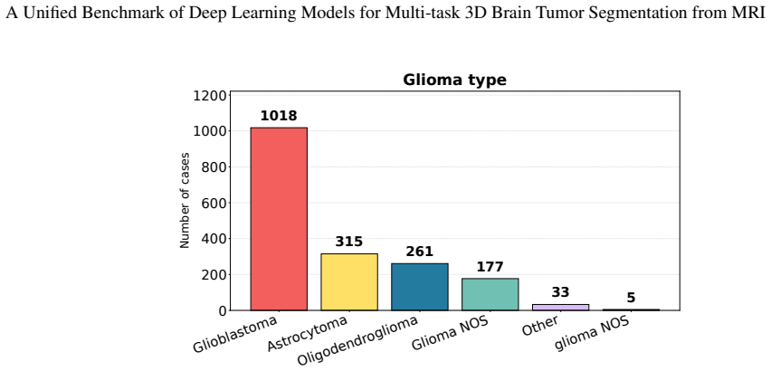
- The two-dataset pattern suggests the state-space advantage is not tied to one tumor type or acquisition scenario, since the ranking holds for both compact meningiomas and distorted post-treatment gliomas.
- SegMambaV2 is the best high-accuracy choice where inference speed matters, while SegMamba offers a lighter-parameter alternative with nearly the same accuracy.
- For hardware-constrained deployment, SegResNet remains the practical mid-point: near-sub-second inference and competitive Dice at a fraction of the parameters of the SSMs and Transformer.
- Swin UNETR's per-sub-region wins indicate that Transformer attention may still be preferable when a specific tumor component is the clinical priority, even if global metrics favor SSMs.
Where Pith is reading between the lines
- Editorial inference: The comparison with challenge leaderboards suggests the local protocol understates what these architectures can do with full training data; an obvious extension is to rerun the exact benchmark using the complete training sets and official validation annotations, which would test whether the SSM advantage persists when more data is available.
- Editorial inference: Because the released SegMamba code omits the FUE module and replaces gated multiplication with addition, and SegMambaV2 approximates its downsampling with a plain convolution, the 'SegMamba family' tested here is really a partially reimplemented variant; a faithful reimplementation could shift the reported gaps.
- Editorial inference: The benchmark uses one crop size, one training budget, and a tenfold lower learning rate for the SSM models; a protocol sweep over these choices would reveal whether the ranking is stable under hyperparameter variation.
- Editorial inference: The per-sub-region results imply that a hybrid architecture — an SSM backbone with a targeted attention module for sub-regions like ET or SNFH — could combine the best of both families; the paper does not test this, but the data suggest it.
Editorial analysis
A structured set of objections, weighed in public.
Referee Report
Summary. The paper reports a unified benchmark of five 3D brain-tumor segmentation architectures—3D U-Net, SegResNet, Swin UNETR, SegMamba, and SegMambaV2—on BraTS 2023 meningioma and BraTS 2024 post-treatment glioma datasets. All models are trained from scratch with a shared MONAI-based preprocessing, augmentation, and evaluation pipeline under five-fold cross-validation, and are compared in terms of Dice, IoU, HD95, inference time, and parameter count. The main claim, stated in the Conclusion, is that the SSM architectures SegMamba and SegMambaV2 consistently achieve the best global performance on both datasets, outperforming the CNN and Transformer baselines.
Significance. If the central claim were supported, this would be a useful contribution: the paper addresses an important problem (heterogeneous evaluation protocols in medical image segmentation), provides code, evaluates two clinically distinct BraTS tasks, includes efficiency metrics alongside accuracy, and documents class imbalance. The five-fold protocol and public benchmark are strengths. However, the main claim is currently not supported as stated, because the evaluated SegMamba and SegMambaV2 implementations differ from the published architectures and were trained with different optimization hyperparameters from the baselines. The benchmark remains valuable as a reproducible comparison of the specific adapted implementations, provided the conclusions are reframed or the confounds are removed.
major comments (3)
- [§3.2.4, §3.2.5, §5] The Conclusion attributes the best results to 'SSM architectures, SegMamba and SegMambaV2,' but the experiments do not evaluate the published SegMamba and SegMambaV2. §3.2.4 states that the SegMamba implementation omits the FUE module, replaces GSC gated multiplication with addition, and uses standard convolutional decoder blocks. §3.2.5 states that SegMambaV2 approximates HS-Downsampling with a single convolution and implements ToOM via axis permutation plus 1D Mamba layers. These are material architectural differences, not cosmetic ones. The observed performance gap may reflect these modifications rather than the SSM design. To support the central claim, the authors should either evaluate the original published implementations or explicitly reframe all conclusions as applying to the adapted implementations, ideally with an ablation showing the modifications do not change relative ranki
- [§3.3, Table 2] The abstract states that all architectures are trained using 'identical ... optimization strategies,' but Table 2 shows that SegMamba and SegMambaV2 use a learning rate of 1e-5 instead of 1e-4 and additionally use gradient clipping with max norm 1. This is a direct confound: the two models that win the comparison were given different optimization settings. The paper offers no control experiment with matched learning rate and clipping for all models, nor evidence that the performance gap is insensitive to these choices. At minimum, the claim of identical optimization must be removed and the results re-interpreted; ideally, the authors should run matched-hyperparameter experiments or a sensitivity analysis.
- [Tables 3 and 4] The global Dice/IoU/HD95 results are reported as five-fold averages without standard deviations, confidence intervals, or significance tests. Some of the gaps supporting the central claim are small (e.g., 0.7559 vs 0.7533 for SegMambaV2 vs SegMamba on BraTS 2024; 0.7245 vs 0.7116 on BraTS 2023). Without variability or a paired test across folds, the statement that SSM architectures 'consistently achieved' the best performance is stronger than the evidence justifies. Report per-fold results and, if possible, a paired significance test or bootstrap confidence intervals.
minor comments (5)
- [Author affiliation] The second author's email contains a duplicated domain: 'luna.hernandez102@ulpgc.es@ulpgc.es'.
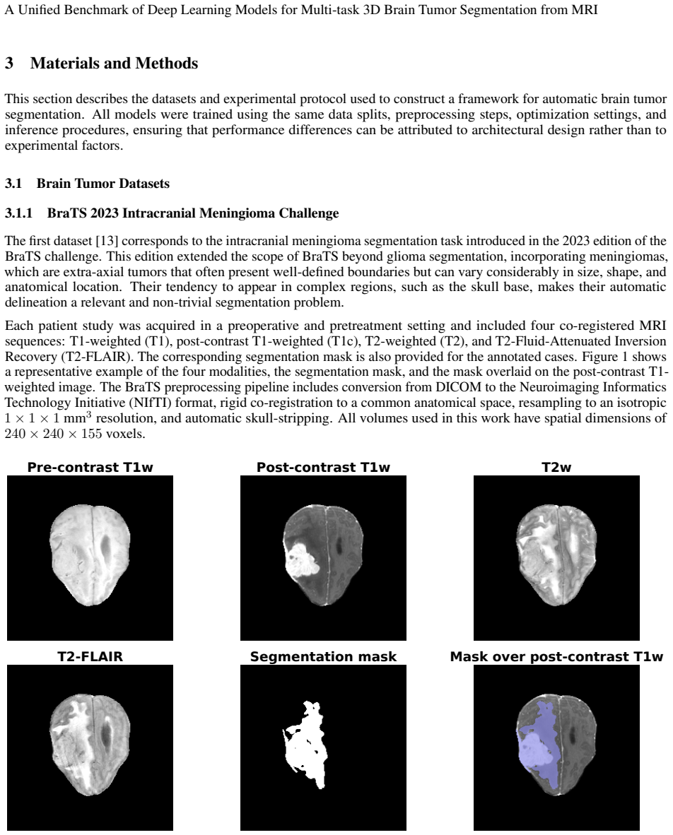
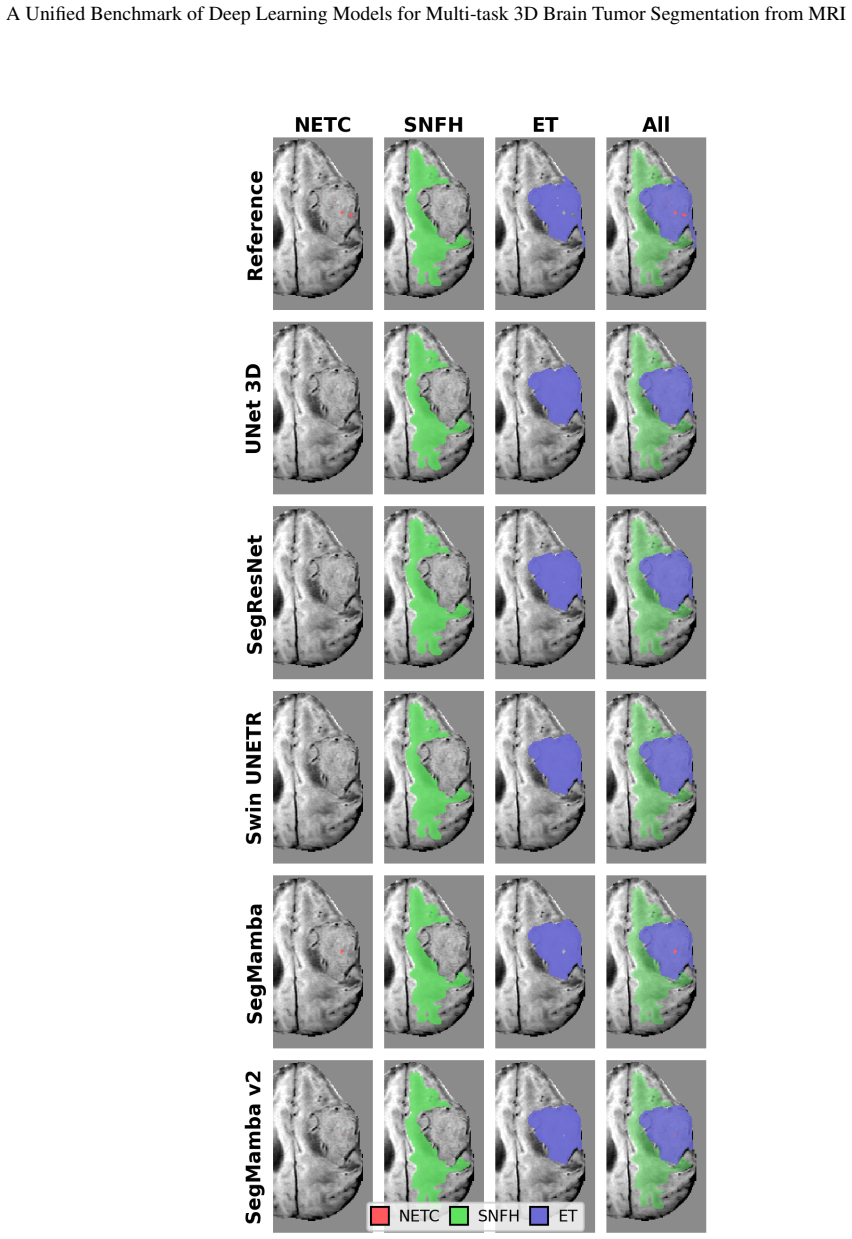
- [Figure 1 and Figure 4] The patient identifier is given as 'BraTS-MEN-00891-000' in Figure 1 and 'BraTS-MEN-00891-00' in Figure 4; please use a consistent identifier.
- [Tables 3 and 4] Use the model name consistently: 'SegMamba V2' in the table header vs 'SegMambaV2' in the text.
- [§3.2.4] The phrase 'the publicly available code differs slightly' understates the listed differences (omitted FUE module, replaced gated multiplication, changed decoder). Suggest describing these as 'substantial differences' or listing them more prominently in a limitations paragraph.
- [§4.5, Tables 7 and 8] The comparison with challenge leaders is clearly labeled as indicative, which is appropriate. However, the table caption 'Best value per column among the models evaluated in this study' is confusing because the table also includes reference methods; consider restructuring the comparison.
Circularity Check
Empirical benchmark; no derivation-level circularity, minor self-citation risk only
full rationale
The paper is an empirical benchmark: it trains five published architectures on two BraTS datasets and reports measured metrics. There is no derivation chain in which a predicted quantity is algebraically equal to, or fitted from, the inputs. The only mild self-citation risk is that the authors' own related-work papers are cited in the introduction and related work, but those citations are not used to compute any result. The central claim that SegMamba/SegMambaV2 achieve the best scores is based on measured Dice, IoU, and HD95 under the stated protocol. The measured scores are therefore not circular in the sense of being equivalent to their inputs by construction. The skeptical concern about altered implementations and different hyperparameters (omitted FUE module, addition in place of gated multiplication, approximated HS-Downsampling, lower learning rate, gradient clipping) is a validity or correctness risk, not a circularity: the results are still measured, not derived from the assumptions. The paper itself explicitly discloses these implementation differences and training deviations (Sections 3.2.4, 3.2.5, and Table 2), which strengthens transparency but does not create circularity. Thus the score is 1, reflecting only the non-load-bearing self-references in related work.
Axiom & Free-Parameter Ledger
free parameters (5)
- Learning rate for SegMamba and SegMambaV2 =
1e-5
- Gradient clipping max norm =
1
- Training crop size =
128x128x128 voxels
- Maximum epochs =
100
- Sliding-window overlap =
25%
axioms (4)
- domain assumption The annotated BraTS training cases are a valid basis for five-fold cross-validation and the resulting metrics generalize to the challenge setting.
- ad hoc to paper The adapted SegMamba and SegMambaV2 code represents the published architectures closely enough to draw conclusions about those architectures.
- standard math MONAI's Dice, IoU, and HD95 implementations measure what the paper claims.
- domain assumption The tumor sub-region labels in BraTS 2023 and BraTS 2024 are correct and consistent.
read the original abstract
Automatic brain tumor segmentation from magnetic resonance imaging (MRI) has become a fundamental task in computer-assisted diagnosis, treatment planning, and disease monitoring. Although numerous deep learning architectures have recently been proposed, objective comparisons remain challenging because published studies often employ different datasets, preprocessing strategies, training protocols, and evaluation procedures. This work presents a unified experimental benchmark for comparing representative convolutional neural networks (CNNs), Transformer-based models, and recent State Space Model (SSM) architectures under homogeneous experimental conditions. Five state-of-the-art three-dimensional segmentation models, including 3D U-Net, SegResNet, Swin UNETR, SegMamba, and SegMambaV2, are evaluated on two brain tumor segmentation datasets representing distinct clinical scenarios: intracranial meningioma segmentation (BraTS 2023) and post-treatment glioma segmentation (BraTS 2024). All architectures are trained using identical preprocessing, data augmentation, optimization strategies, and evaluation protocols to ensure a fair comparison. Performance is assessed using segmentation accuracy metrics together with computational cost indicators, including inference time and the size of each model. The results provide practical insights into the trade-offs between segmentation accuracy and computational efficiency, highlighting the suitability of different architectural paradigms for challenging three-dimensional brain tumor segmentation tasks.
Figures






Reference graph
Works this paper leans on
-
[1]
U-Net: Convolutional networks for biomedical image segmentation
Olaf Ronneberger, Philipp Fischer, and Thomas Brox. U-Net: Convolutional networks for biomedical image segmentation. In Nassir Navab, Joachim Hornegger, William M. Wells, and Alejandro F. Frangi, editors,Medical Image Computing and Computer-Assisted Intervention – MICCAI 2015, pages 234–241, Cham, 2015. Springer International Publishing
2015
-
[2]
3D U-Net: Learning dense volumetric segmentation from sparse annotation
Özgün Çiçek, Ahmed Abdulkadir, Soeren S Lienkamp, Thomas Brox, and Olaf Ronneberger. 3D U-Net: Learning dense volumetric segmentation from sparse annotation. InInternational Conference on Medical Image Computing and Computer-Assisted Intervention (MICCAI), pages 424–432. Springer, 2016. 23 A Unified Benchmark of Deep Learning Models for Multi-task 3D Brai...
2016
-
[3]
Roth, and Daguang Xu
Ali Hatamizadeh, Yucheng Tang, Vishwesh Nath, Dong Yang, Andriy Myronenko, Bennett Landman, Holger R. Roth, and Daguang Xu. UNETR: Transformers for 3D medical image segmentation. In2022 IEEE/CVF Winter Conference on Applications of Computer Vision (WACV), pages 1748–1758, 2022
2022
-
[4]
Roth, and Daguang Xu
Ali Hatamizadeh, Vishwesh Nath, Yucheng Tang, Dong Yang, Holger R. Roth, and Daguang Xu. Swin UNETR: Swin transformers for semantic segmentation of brain tumors in MRI images. In Alessandro Crimi and Spyridon Bakas, editors,Brainlesion: Glioma, Multiple Sclerosis, Stroke and Traumatic Brain Injuries, pages 272–284, Cham, 2022. Springer International Publishing
2022
-
[5]
Efficiently modeling long sequences with structured state spaces
Albert Gu, Karan Goel, and Christopher Re. Efficiently modeling long sequences with structured state spaces. In International Conference on Learning Representations, 2022
2022
-
[6]
Mamba: Linear-time sequence modeling with selective state spaces
Albert Gu and Tri Dao. Mamba: Linear-time sequence modeling with selective state spaces. InFirst conference on language modeling, 2024
2024
-
[7]
U-Mamba: Enhancing long-range dependency for biomedical image segmentation
Jun Ma, Feifei Li, and Bo Wang. U-Mamba: Enhancing long-range dependency for biomedical image segmentation. arXiv preprint arXiv:2401.04722, 2024
Pith/arXiv arXiv 2024
-
[8]
VM-UNet: Vision Mamba UNet for medical image segmenta- tion.ACM Transactions on Multimedia Computing, Communications and Applications, 2024
Jiacheng Ruan, Jincheng Li, and Suncheng Xiang. VM-UNet: Vision Mamba UNet for medical image segmenta- tion.ACM Transactions on Multimedia Computing, Communications and Applications, 2024
2024
-
[9]
Zhaohu Xing, Tian Ye, Yijun Yang, Guang Liu, and Lei Zhu. SegMamba: Long-range sequential modeling Mamba for 3D medical image segmentation.arXiv preprint arXiv:2401.13560, 2024
Pith/arXiv arXiv 2024
-
[10]
SegMamba-V2: Long-range sequential modeling Mamba for general 3-D medical image segmentation.IEEE Transactions on Medical Imaging, 2025
Zhaohu Xing, Tian Ye, Yijun Yang, Du Cai, Baowen Gai, Xiao-Jian Wu, Feng Gao, and Lei Zhu. SegMamba-V2: Long-range sequential modeling Mamba for general 3-D medical image segmentation.IEEE Transactions on Medical Imaging, 2025
2025
-
[11]
3D MRI brain tumor segmentation using autoencoder regularization
Andriy Myronenko. 3D MRI brain tumor segmentation using autoencoder regularization. In Alessandro Crimi, Spyridon Bakas, Hugo Kuijf, Farahani Keyvan, Mauricio Reyes, and Theo van Walsum, editors,Brainlesion: Glioma, Multiple Sclerosis, Stroke and Traumatic Brain Injuries, pages 311–320, Cham, 2019. Springer Interna- tional Publishing
2019
-
[12]
Bjoern H. Menze, Andras Jakab, Stefan Bauer, Jayashree Kalpathy-Cramer, Keyvan Farahani, Justin Kirby, Yuliya Burren, Nicole Porz, Johannes Slotboom, Roland Wiest, Levente Lanczi, Elizabeth Gerstner, Marc-André Weber, Tal Arbel, Brian B. Avants, Nicholas Ayache, Patricia Buendia, D. Louis Collins, Nicolas Cordier, Jason J. Corso, Antonio Criminisi, Tilak ...
1993
-
[13]
The ASNR-MICCAI brain tumor segmentation (BraTS) challenge 2023: Intracranial meningioma, 2023
Dominic LaBella, Maruf Adewole, Michelle Alonso-Basanta, Talissa Altes, Syed Muhammad Anwar, Ujjwal Baid, Timothy Bergquist, Radhika Bhalerao, Sully Chen, Verena Chung, Gian-Marco Conte, Farouk Dako, James Eddy, Ivan Ezhov, Devon Godfrey, Fathi Hilal, Ariana Familiar, Keyvan Farahani, Juan Eugenio Iglesias, Zhifan Jiang, Elaine Johanson, Anahita Fathi Kaz...
2023
-
[14]
Maria Correia de Verdier, Rachit Saluja, Louis Gagnon, Dominic LaBella, Ujjwall Baid, Nourel Hoda Tahon, Martha Foltyn-Dumitru, Jikai Zhang, Maram Alafif, and Saif et al. Baig. The 2024 brain tumor segmentation (BraTS) challenge: Glioma segmentation on post-treatment MRI. Technical report, Cornell Univeristy, 2024
2024
-
[15]
Performance of machine learning algorithms for glioma segmentation of brain MRI: a systematic literature review and meta-analysis.European Radiology, 31(12):9638–9653, 2021
Evi J van Kempen, Max Post, Manoj Mannil, Richard L Witkam, Mark Ter Laan, Ajay Patel, Frederick JA Meijer, and Dylan Henssen. Performance of machine learning algorithms for glioma segmentation of brain MRI: a systematic literature review and meta-analysis.European Radiology, 31(12):9638–9653, 2021
2021
-
[16]
A review on deep learning methods for glioma segmentation, limitations, and future perspectives.Journal of Imaging, 11(8):269, 2025
Cecilia Diana-Albelda, Álvaro García-Martín, and Jesus Bescos. A review on deep learning methods for glioma segmentation, limitations, and future perspectives.Journal of Imaging, 11(8):269, 2025. 24 A Unified Benchmark of Deep Learning Models for Multi-task 3D Brain Tumor Segmentation from MRI
2025
-
[17]
A review of deep learning for brain tumor analysis in MRI.NPJ Precision Oncology, 9(1):2, 2025
Felix J Dorfner, Jay B Patel, Jayashree Kalpathy-Cramer, Elizabeth R Gerstner, and Christopher P Bridge. A review of deep learning for brain tumor analysis in MRI.NPJ Precision Oncology, 9(1):2, 2025
2025
-
[18]
Advanced deep learning and machine learning techniques for MRI brain tumor analysis: A review.Sensors, 25(9), 2025
Rim Missaoui, Wided Hechkel, Wajdi Saadaoui, Abdelhamid Helali, and Marco Leo. Advanced deep learning and machine learning techniques for MRI brain tumor analysis: A review.Sensors, 25(9), 2025
2025
-
[19]
Fully convolutional networks for semantic segmentation
Jonathan Long, Evan Shelhamer, and Trevor Darrell. Fully convolutional networks for semantic segmentation. In 2015 IEEE Conference on Computer Vision and Pattern Recognition (CVPR), pages 3431–3440, 2015
2015
-
[20]
Attention U-Net: Learning where to look for the pancreas
Ozan Oktay, Jo Schlemper, Loic Le Folgoc, Matthew Lee, Mattias Heinrich, Kazunari Misawa, Kensaku Mori, Steven McDonagh, Nils Y Hammerla, Bernhard Kainz, Ben Glocker, and Daniel Rueckert. Attention U-Net: Learning where to look for the pancreas. Technical report, Cornell University, 2018
2018
-
[21]
nnU-Net: a self- configuring method for deep learning-based biomedical image segmentation.Nature methods, 18(2):203–211, 2021
Fabian Isensee, Paul F Jaeger, Simon AA Kohl, Jens Petersen, and Klaus H Maier-Hein. nnU-Net: a self- configuring method for deep learning-based biomedical image segmentation.Nature methods, 18(2):203–211, 2021
2021
-
[22]
Performance of convolutional neural networks for the classification of brain tumors using magnetic resonance imaging.Heliyon, 10(3), 2024
Daniel Reyes and Javier Sánchez. Performance of convolutional neural networks for the classification of brain tumors using magnetic resonance imaging.Heliyon, 10(3), 2024
2024
-
[23]
High accuracy brain tumor classification with EfficientNet and magnetic resonance images
Juan Manuel Medina and Javier Sánchez. High accuracy brain tumor classification with EfficientNet and magnetic resonance images. InProceedings of the 5th International Conference on Advances in Signal Processing and Artificial Intelligence (ASPAI 2023), pages 151–156, Tenerife, Canary Islands, Spain, 2023. International Frequency Sensor Association (IFSA)...
2023
-
[24]
Gomez, Łukasz Kaiser, and Illia Polosukhin
Ashish Vaswani, Noam Shazeer, Niki Parmar, Jakob Uszkoreit, Llion Jones, Aidan N. Gomez, Łukasz Kaiser, and Illia Polosukhin. Attention is all you need. InAdvances in Neural Information Processing Systems (NeurIPS), volume 30, 2017
2017
-
[25]
Alexey Dosovitskiy, Lucas Beyer, Alexander Kolesnikov, Dirk Weissenborn, Xiaohua Zhai, Thomas Unterthiner, Mostafa Dehghani, Matthias Minderer, Georg Heigold, Sylvain Gelly, et al. An image is worth 16x16 words: Transformers for image recognition at scale.arXiv preprint arXiv:2010.11929, 2020
Pith/arXiv arXiv 2010
-
[26]
TransUNet: Rethinking the U-Net architecture design for medical image segmentation through the lens of transformers.Medical Image Analysis, page 103280, 2024
Jieneng Chen, Jieru Mei, Xianhang Li, Yongyi Lu, Qihang Yu, Qingyue Wei, Xiangde Luo, Yutong Xie, Ehsan Adeli, Yan Wang, et al. TransUNet: Rethinking the U-Net architecture design for medical image segmentation through the lens of transformers.Medical Image Analysis, page 103280, 2024
2024
-
[27]
TransBTS: Multimodal brain tumor segmentation using transformer
Wenxuan Wang, Chen Chen, Meng Ding, Hong Yu, Sen Zha, and Jiangyun Li. TransBTS: Multimodal brain tumor segmentation using transformer. In Marleen de Bruijne, Philippe C. Cattin, Stéphane Cotin, Nicolas Padoy, Stefanie Speidel, Yefeng Zheng, and Caroline Essert, editors,Medical Image Computing and Computer Assisted Intervention – MICCAI 2021, pages 109–11...
2021
-
[28]
Swin transformer: Hierarchical vision transformer using shifted windows
Ze Liu, Yutong Lin, Yue Cao, Han Hu, Yixuan Wei, Zheng Zhang, Stephen Lin, and Baining Guo. Swin transformer: Hierarchical vision transformer using shifted windows. InProceedings of the IEEE/CVF international conference on computer vision, pages 10012–10022, 2021
2021
-
[29]
Swin transformer V2: Scaling up capacity and resolution
Ze Liu, Han Hu, Yutong Lin, Zhuliang Yao, Zhenda Xie, Yixuan Wei, Jia Ning, Yue Cao, Zheng Zhang, Li Dong, et al. Swin transformer V2: Scaling up capacity and resolution. InProceedings of the IEEE/CVF conference on computer vision and pattern recognition, pages 12009–12019, 2022
2022
-
[30]
Evaluation of vision transformers for multi-organ tumor classification using MRI and CT imaging.Electronics, 14(15):2976, 2025
Óscar A Martín and Javier Sánchez. Evaluation of vision transformers for multi-organ tumor classification using MRI and CT imaging.Electronics, 14(15):2976, 2025
2025
-
[31]
Vision Mamba: efficient visual representation learning with bidirectional state space model
Lianghui Zhu, Bencheng Liao, Qian Zhang, Xinlong Wang, Wenyu Liu, and Xinggang Wang. Vision Mamba: efficient visual representation learning with bidirectional state space model. InProceedings of the 41st International Conference on Machine Learning, ICML’24. JMLR.org, 2024
2024
-
[32]
VMamba: visual state space model
Yue Liu, Yunjie Tian, Yuzhong Zhao, Hongtian Yu, Lingxi Xie, Yaowei Wang, Qixiang Ye, Jianbin Jiao, and Yunfan Liu. VMamba: visual state space model. InProceedings of the 38th International Conference on Neural Information Processing Systems, NIPS ’24, Red Hook, NY , USA, 2024. Curran Associates Inc
2024
-
[33]
VM-UNET-V2: Rethinking vision Mamba UNet for medical image segmentation
Mingya Zhang, Yue Yu, Sun Jin, Limei Gu, Tingsheng Ling, and Xianping Tao. VM-UNET-V2: Rethinking vision Mamba UNet for medical image segmentation. InBioinformatics Research and Applications: 20th International Symposium, ISBRA 2024, Kunming, China, July 19–21, 2024, Proceedings, Part I, page 335–346, Berlin, Heidelberg, 2024. Springer-Verlag
2024
-
[34]
Mamba-UNet: UNet-like pure visual Mamba for medical image segmentation
Ziyang Wang, Jian-Qing Zheng, Yichi Zhang, Ge Cui, and Lei Li. Mamba-UNet: UNet-like pure visual Mamba for medical image segmentation. Technical report, Cornell University, 2024. 25 A Unified Benchmark of Deep Learning Models for Multi-task 3D Brain Tumor Segmentation from MRI
2024
-
[35]
Swin-UMamba: Mamba-based UNet with ImageNet-based pretraining
Jiarun Liu, Hao Yang, Hong-Yu Zhou, Yan Xi, Lequan Yu, Cheng Li, Yong Liang, Guangming Shi, Yizhou Yu, Shaoting Zhang, Hairong Zheng, and Shanshan Wang. Swin-UMamba: Mamba-based UNet with ImageNet-based pretraining . InProceedings of Medical Image Computing and Computer Assisted Intervention – MICCAI 2024, volume LNCS 15009. Springer Nature Switzerland, O...
2024
-
[36]
Chen, and Jian Wu
Jinhong Wang, Jintai Chen, Danny Z. Chen, and Jian Wu. LKM-UNet: Large kernel vision Mamba UNet for medical image segmentation. InProceedings of Medical Image Computing and Computer Assisted Intervention – MICCAI 2024, volume LNCS 15008. Springer Nature Switzerland, October 2024
2024
-
[37]
Rotate to scan: UNet-like Mamba with triplet SSM module for medical image segmentation
Hao Tang, Lianglun Cheng, Guoheng Huang, Zhengguang Tan, Junhao Lu, and Kaihong Wu. Rotate to scan: UNet-like Mamba with triplet SSM module for medical image segmentation. Technical report, Cornell University, 2024
2024
-
[38]
Computation-efficient era: A comprehensive survey of state space models in medical image analysis
Moein Heidari, Sina Ghorbani Kolahi, Sanaz Karimijafarbigloo, Bobby Azad, Afshin Bozorgpour, Soheila Hatami, Reza Azad, Ali Diba, Ulas Bagci, Dorit Merhof, and Ilker Hacihaliloglu. Computation-efficient era: A comprehensive survey of state space models in medical image analysis. Technical report, Cornell University, 2024
2024
-
[39]
A comprehensive survey of Mamba architectures for medical image analysis: Classification, segmentation, restoration and beyond
Shubhi Bansal, Sreeharish A, Madhava Prasath J, Manikandan S, Sreekanth Madisetty, Mohammad Zia Ur Rehman, Chandravardhan Singh Raghaw, Gaurav Duggal, and Nagendra Kumar. A comprehensive survey of Mamba architectures for medical image analysis: Classification, segmentation, restoration and beyond. Technical report, Cornell University, 2025
2025
-
[40]
Mamba in vision: A comprehensive survey of techniques and applications
Md Maklachur Rahman, Abdullah Aman Tutul, Ankur Nath, Lamyanba Laishram, Soon Ki Jung, and Tracy Hammond. Mamba in vision: A comprehensive survey of techniques and applications. Technical report, Cornell University, 2024
2024
-
[41]
Advancing the cancer genome atlas glioma MRI collections with expert segmentation labels and radiomic features.Scientific Data, 4(1):170117, 2017
Spyridon Bakas, Hamed Akbari, Aristeidis Sotiras, Michel Bilello, Martin Rozycki, Justin S Kirby, John B Freymann, Keyvan Farahani, and Christos Davatzikos. Advancing the cancer genome atlas glioma MRI collections with expert segmentation labels and radiomic features.Scientific Data, 4(1):170117, 2017
2017
-
[42]
Shah, Blaise V
Anahita Fathi Kazerooni, Nastaran Khalili, Xinyang Liu, Debanjan Haldar, Zhifan Jiang, Anna Zapaishchykova, Julija Pavaine, Lubdha M. Shah, Blaise V . Jones, Nakul Sheth, Sanjay P. Prabhu, Aaron S. McAllister, Wenxin Tu, Khanak K. Nandolia, Andres F. Rodriguez, Ibraheem Salman Shaikh, Mariana Sanchez-Montano, Hollie Anne Lai, Maruf Adewole, Jake Albrecht,...
2023
-
[43]
Metrics reloaded: recommendations for image analysis validation.Nature methods, 21(2):195–212, 2024
Lena Maier-Hein, Annika Reinke, Patrick Godau, Minu D Tizabi, Florian Buettner, Evangelia Christodoulou, Ben Glocker, Fabian Isensee, Jens Kleesiek, Michal Kozubek, et al. Metrics reloaded: recommendations for image analysis validation.Nature methods, 21(2):195–212, 2024
2024
-
[44]
A multi-institutional meningioma MRI dataset for automated multi-sequence image segmentation.Scientific Data, 11(1):496, 2024
Dominic LaBella, Omaditya Khanna, Shan McBurney-Lin, Ryan Mclean, Pierre Nedelec, Arif S Rashid, Nourel Hoda Tahon, Talissa Altes, Ujjwal Baid, Radhika Bhalerao, et al. A multi-institutional meningioma MRI dataset for automated multi-sequence image segmentation.Scientific Data, 11(1):496, 2024
2024
-
[45]
MONAI: An open-source framework for deep learning in healthcare
M Jorge Cardoso, Wenqi Li, Richard Brown, Nic Ma, Eric Kerfoot, Yiheng Wang, Benjamin Murrey, Andriy Myronenko, Can Zhao, Dong Yang, et al. MONAI: An open-source framework for deep learning in healthcare. arXiv preprint arXiv:2211.02701, 2022
Pith/arXiv arXiv 2022
-
[46]
V-Net: Fully convolutional neural networks for volumetric medical image segmentation
Fausto Milletari, Nassir Navab, and Seyed-Ahmad Ahmadi. V-Net: Fully convolutional neural networks for volumetric medical image segmentation. In2016 fourth international conference on 3D vision (3DV), pages 565–571. IEEE, 2016
2016
-
[47]
Decoupled weight decay regularization
Ilya Loshchilov and Frank Hutter. Decoupled weight decay regularization. InInternational Conference on Learning Representations, 2019
2019
-
[48]
Auto3DSeg: Automated 3D medical image segmentation.arXiv preprint, 2023
Yufan He, Dong Yang, Andriy Myronenko, Daguang Xu, et al. Auto3DSeg: Automated 3D medical image segmentation.arXiv preprint, 2023. 26 A Unified Benchmark of Deep Learning Models for Multi-task 3D Brain Tumor Segmentation from MRI
2023
-
[49]
Improved multi-task brain tumour segmentation with synthetic data augmentation, 2024
André Ferreira, Tiago Jesus, Behrus Puladi, Jens Kleesiek, Victor Alves, and Jan Egger. Improved multi-task brain tumour segmentation with synthetic data augmentation, 2024. 27
2024
discussion (0)
Sign in with ORCID, Apple, or X to comment. Anyone can read and Pith papers without signing in.