REVIEW 3 major objections 5 minor 82 references
A single-pass latent transport network can synthesize breast DCE-MRI contrast enhancement at any acquisition time from the pre-contrast scan alone, matching real scans closely enough to leave clinical management unchanged in 70% of reader-s
Reviewed by Pith at T0; open to challenge. T0 means a machine referee read the full paper against a public rubric. the ladder, T0–T4 →
T0 review · deepseek-v4-flash
2026-08-03 07:52 UTC pith:NPJ3UYPI
load-bearing objection A thorough empirical package for latent contrast synthesis with a real clinical validation stack; the central continuous-time claim is untested and the abstract overstates the metric coverage. the 3 major comments →
Dense Temporal Contrast Synthesis via Conditioned Latent Transport
The pith
A machine-rendered reading of the paper's core claim, the machinery that carries it, and where it could break.
Core claim
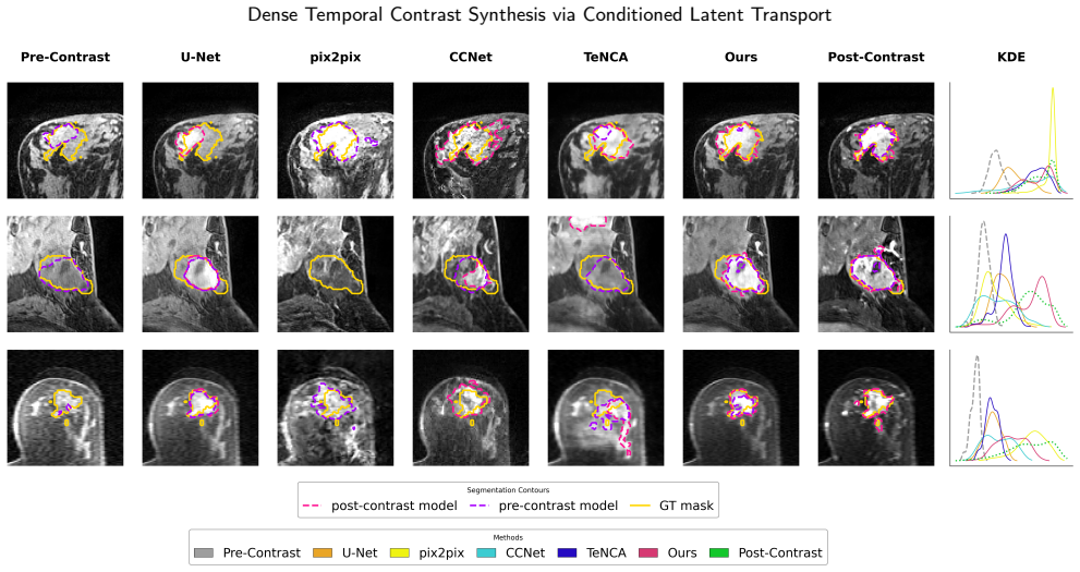
The central discovery is that contrast enhancement in breast DCE-MRI can be treated as a residual in latent space: the enhancement map Δz = z_post − z_pre, conditioned on a continuous acquisition time τ, contains almost all the clinically relevant information. The model anchors the generative trajectory to the pre-contrast latent, predicts Δz from a noisy interpolated intermediate state, and reconstructs the enhanced image in one pass, preserving static anatomy while generating smooth, patient-specific kinetics. The authors support this with gains across spatial, perceptual, temporal, and distributional metrics, external-cohort generalization, improved downstream tumor segmentation, and a fo
What carries the argument
The conditioned latent transport network: a frozen autoencoder with 4× spatial downsampling maps images into a high-fidelity latent space; a U-Net receives the noisy interpolated latent concatenated with the pre-contrast anchor, with sinusoidal acquisition-time embedding τ injected via adaptive group normalization. The network is trained to regress the constant latent subtraction map Δz = z_post − z_pre, using MSE, LPIPS, and focal-frequency losses. At inference, a single pass with one fixed patient-level noise map yields the enhanced latent, decoded by the frozen decoder. The load-bearing design is that τ is continuous and decoupled from the degradation schedule, and anatomy never has to be
Load-bearing premise
The paired pre- and post-contrast slices are assumed to be adequately aligned so that the learned latent difference Δz represents true contrast uptake; the paper only qualitatively assessed registration and did not quantify or correct patient motion between acquisitions.
What would settle it
Compute the same downstream segmentation and temporal metrics after applying strict rigid or deformable motion correction to every pre/post pair. If the 22.4% Dice improvement and low temporal error largely disappear, the claimed enhancement signal was partly an alignment artifact rather than synthesized physiology.
If this is right
- Contrast-free or contrast-reduced breast MRI becomes technically plausible: for 70% of evaluated cases, synthetic scans would not change management decisions.
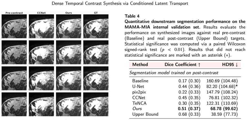
- Downstream tumor segmentation on synthetic images approaches the real post-contrast upper bound (Dice 0.60 vs 0.63 with a pre-contrast-trained network), reducing boundary error by over 39%.
- Continuous time conditioning lets clinicians query enhancement at arbitrary acquisition times, enabling pharmacokinetic curve analysis without additional scan phases.
- The method remains the best generative baseline on an unseen external cohort despite faster wash-in dynamics and different scanner noise, indicating some tolerance to protocol shifts.
- Single-pass inference avoids iterative sampling, making synthesis practical within clinical time budgets.
Where Pith is reading between the lines
- If the latent subtraction truly isolates physiology from anatomy, the same transport could map low-dose or early-phase contrast to full enhancement, turning the framework into a dose-reduction tool rather than a contrast-elimination one.
- The decoupling of physical time τ from the degradation schedule invites a natural extension: replace linear interpolation with an explicit physiological pharmacokinetic model as the interpolant, which the paper itself flags as future work.
- The patient-level fixed noise yields cheap Monte Carlo uncertainty maps; a formal calibration study could turn the reader-observed 49% informative rate into a quantitative safety guarantee.
- The finding that paired synthetic-vs-real disagreement is smaller than inter-reader disagreement sets a target for other synthesis methods: clinical equivalence, not perfect pixels, is the bar.
Editorial analysis
A structured set of objections, weighed in public.
Referee Report
Summary. The paper proposes a conditioned latent transport framework for synthesizing post-contrast breast DCE-MRI from pre-contrast images. A frozen VAE compresses images into a latent space; a U-Net is trained to predict the residual Δz = z_post − z_pre from a noisy interpolation between z_pre and z_post, conditioned on a sinusoidal embedding of the physical acquisition time τ. At inference, the model produces a target phase in a single forward pass using a fixed patient-level noise map. The method is evaluated on an internal cohort (MAMA-MIA), an external cohort (Karolinska), compared against U-Net, pix2pix, CCNet, and TeNCA via spatial, perceptual, distributional, and temporal metrics, and further assessed by downstream tumor segmentation and a four-radiologist reader study. The central claims are: (1) continuous-time synthesis at any τ, (2) state-of-the-art quantitative performance, (3) robustness to domain shift, (4) improved segmentation, and (5) clinical viability in 70% of reader-study cases.
Significance. If the central claims hold, the work is significant: it targets a clinically important problem (reducing GBCA exposure in breast MRI) and combines a deterministic single-step generative model with an unusually thorough evaluation protocol — external cohort, downstream task, reader study, and ablations. The pre-contrast anchoring and residual prediction are sensible inductive biases, and the extensive validation framework sets a good example for the field. The segmentation and reader-study results are genuinely evaluated on held-out data and are the strongest part of the paper. However, the headline claim of continuous-time synthesis is not quantitatively validated, and the abstract overstates metric superiority. These issues must be resolved before the paper can be accepted.
major comments (3)
- [§3.1, §3.5, Table 1, Appendix D] The central claim of continuous-time synthesis at arbitrary τ is not tested. All temporal metrics (PTE, DTW, DTW-ROI) in Table 1 and the external rows are computed on the same discrete post-contrast phases used as training targets. There is no held-out τ experiment, no evaluation at an interpolated time point, and no ablation that varies τ continuously while measuring accuracy. The model is trained as a pointwise regressor from (z_pre, τ) to z_post; the forward corruption schedule t is decoupled from τ, and Appendix D shows that an explicit attempt to add continuous supervision via interpolated latents (Table D.7) worsened PTE from 44.57 to 60.79 and was abandoned. Thus the paper's title, abstract, and §3.1 claim that the model 'synthesizes patient-specific contrast evolution at any acquisition time' is unsupported by quantitative evidence. Please either add a held-out-phase evaluation (
- [Abstract and §5.1, Table 1] The abstract states that the method 'outperforms baseline and the state-of-the-art models across spatial, perceptual, temporal, and distributional metrics.' This is contradicted by Table 1: on SSIM (a spatial metric) the method scores 0.71 versus 0.73 for both pix2pix and TeNCA on the internal validation set, and on FRD (a distributional radiomic metric) pix2pix scores 4.50 vs. the proposed method's 4.98. The explanations in §5.1 about 'algorithmic biases' of the baselines are interpretive and do not change the metric values. The abstract and any summary statements should be revised to say 'most metrics' or explicitly acknowledge these two exceptions, so that readers are not misled about the scope of the improvement.
- [§6 (Limitations), §3.3–3.4] The method's physiological interpretation rests on the assumption that Δz = z_post − z_pre represents true contrast enhancement. Section 6 states that pre- and post-contrast registration was 'only qualitatively assessed' and that patient motion 'was not strictly quantified or corrected.' If motion between the pre- and post-contrast acquisitions is substantial, the residual target is corrupted by misalignment, and both the temporal metrics (which are computed on the mean intensity inside the ground-truth mask) and the downstream segmentation improvements could partly reflect alignment artifacts rather than genuine contrast uptake. Because this is a core assumption of the latent transport formulation, please provide a quantitative motion analysis (e.g., displacement estimates within the tumor region) or a controlled experiment with motion correction, or at least a discussion of the expecte
minor comments (5)
- [Table 3 and §5.3.2] The ablation table reports FID-Dinov2 values of 141.87 (w/o pre-conditioning) and 154.56 (w pre-conditioning), while the text gives 141.91 and 155.17. Please make the numbers consistent.
- [§5.5] Typo: 'an strong preference' should be 'a strong preference.'
- [§4.2] The metric is first called 'Time-to-Peak Error (PTE)' and then defined as 'Peak Timing Error (PTE)'. The equation uses 'TTPE'. Please unify the abbreviation.
- [§4.2 and Table 1] It would be helpful to state clearly that the temporal metrics for pix2pix are omitted because the model is not conditioned on acquisition time; currently this is explained in §4.3 but not in the metric section. Consider adding a note near Table 1.
- [§5.3.1] The ablation description in the text says the addition of stochastic regularization 'injects essential micro-textural realism' and improves FID-Dinov2 to 145.06, but the final model with Fourier loss has FID-Dinov2 154.56, which is worse. The ordering of the ablation rows is non-monotonic; please verify the row labels and the narrative.
Circularity Check
No significant circularity; the central predictions are not defined by their inputs, and the self-citations are limited to benchmarks/comparators rather than load-bearing derivation steps.
full rationale
The paper's core derivation is a supervised latent regression: the network maps (z_pre, tau) to a residual Delta_z = z_post - z_pre, reconstructs z_post = z_pre + Delta_z, and is evaluated on held-out cases. No equation in the paper defines a reported result in terms of a fitted parameter or renames a fit as a prediction. The residual parameterization is a prediction target, not a fitted input, and adding z_pre back is simple bookkeeping. The downstream segmentation evaluation uses independently trained nnUNet models on real images, the external KI cohort provides an out-of-distribution test, and the reader study is a blinded human evaluation; these are independent forms of evidence rather than assumptions built into the synthesis objective. The self-cited works (TeNCA, CCNet, FRD, MAMA-MIA) are used as comparators, metrics, or data sources, and none is invoked as a uniqueness theorem or as the justification for the method's central formulation. The paper itself acknowledges the main weaknesses: registration was only qualitatively assessed, and the attempted temporal sampling strategy for sparse early/late acquisition times increased PTE (Table D.7) and was abandoned. The continuous-time claim is indeed not quantitatively tested at held-out or interpolated tau values, since all temporal metrics are computed on the discrete phases that served as training targets; however, this is an unsupported validation claim, not a circular derivation. For these reasons, the appropriate finding is no significant circularity, with a low score reflecting the presence of several non-load-bearing self-citations in the evaluation setup.
Axiom & Free-Parameter Ledger
free parameters (3)
- Loss weights λ_MSE, λ_LPIPS, λ_FFL =
1, 5, 50
- Noise scale σ_noise =
0.1
- VAE latent scaling factor =
1.0259
axioms (4)
- domain assumption Paired pre- and post-contrast images are spatially aligned after preprocessing
- domain assumption Contrast enhancement is a deterministic function of pre-contrast anatomy and acquisition time τ
- ad hoc to paper Linear latent interpolation between z_pre and z_post approximates the pharmacokinetic trajectory
- domain assumption The frozen VAE latent space preserves clinically relevant high-frequency detail
read the original abstract
Dynamic contrast-enhanced magnetic resonance imaging (DCE-MRI) is essential for breast cancer management, but reliance on gadolinium-based contrast agents (GBCAs) restricts use in contraindicated populations, prolongs scan protocols, and presents environmental toxicity concerns. Contrast synthesis offers a non-invasive alternative; however, existing approaches struggle to balance spatial realism with temporal continuity, suffer from slow iterative sampling, underutilize structural priors, and lack clinical validation. We propose a novel conditioned latent transport framework that predicts contrast enhancement in a single forward pass. By anchoring the latent trajectory to the pre-contrast anatomy and applying continuous time conditioning, the model synthesizes patient-specific contrast evolution at any acquisition time. The proposed approach outperforms baseline and the state-of-the-art models across spatial, perceptual, temporal, and distributional metrics. Evaluated on an independent external cohort, the method demonstrates robustness to domain shifts induced by scanner noise as well as differing acquisition protocol. Furthermore, our synthetic contrast enhancement significantly improved downstream tumor segmentation performance, yielding a 22.4% relative increase in Dice coefficient (0.60 vs. 0.49 baseline pre-contrast, p < 0.01), reducing boundary segmentation error by over 39%, while outperforming all other generative model baselines. Finally, a reader study involving four breast radiologists evaluated the image quality, kinetic fidelity, and diagnostic viability of our synthesized sequences across 40 randomly selected cases. The results demonstrated that in 70% of cases, synthesized images provided sufficient clinical information to support the same management decisions as real DCE-MRI, suggesting a path toward safer and faster contrast-free or contrast-reduced imaging workflows.
Figures



Reference graph
Works this paper leans on
-
[1]
BioMed research international , volume=
Role of magnetic resonance imaging in the preoperative staging and work-up of patients affected by invasive lobular carcinoma or invasive ductolobular carcinoma , author=. BioMed research international , volume=. 2018 , publisher=
2018
-
[2]
The breast journal , volume=
Breast MRI in invasive lobular carcinoma: a useful investigation in surgical planning? , author=. The breast journal , volume=. 2016 , publisher=
2016
-
[3]
Radiology , volume=
Impact of preoperative breast MR imaging and MR-guided surgery on diagnosis and surgical outcome of women with invasive breast cancer with and without DCIS component , author=. Radiology , volume=. 2017 , publisher=
2017
-
[4]
World journal of surgery , volume=
Preoperative MRI of the breast (POMB) influences primary treatment in breast cancer: a prospective, randomized, multicenter study , author=. World journal of surgery , volume=. 2014 , publisher=
2014
-
[5]
Radiology , volume=
Dynamic breast MR imaging: are signal intensity time course data useful for differential diagnosis of enhancing lesions? , author=. Radiology , volume=. 1999 , publisher=
1999
-
[6]
Academic radiology , volume=
Breast DCE-MRI: influence of postcontrast timing on automated lesion kinetics assessments and discrimination of benign and malignant lesions , author=. Academic radiology , volume=. 2014 , publisher=
2014
-
[7]
Journal of British Surgery , volume=
Association between underestimation of tumour size by imaging and incomplete excision in breast-conserving surgery for breast cancer , author=. Journal of British Surgery , volume=. 2016 , publisher=
2016
-
[8]
Breast cancer research and treatment , volume=
Comparison of magnetic resonance imaging, multidetector row computed tomography, ultrasonography, and mammography for tumor extension of breast cancer , author=. Breast cancer research and treatment , volume=. 2008 , publisher=
2008
-
[9]
CA: a cancer journal for clinicians , volume=
American Cancer Society guidelines for breast screening with MRI as an adjunct to mammography , author=. CA: a cancer journal for clinicians , volume=. 2007 , publisher=
2007
-
[10]
Journal of the American College of Radiology , volume=
Breast cancer screening in women at higher-than-average risk: recommendations from the ACR , author=. Journal of the American College of Radiology , volume=. 2018 , publisher=
2018
-
[11]
Radiology , volume=
Breast MR imaging screening in women with a history of breast conservation therapy , author=. Radiology , volume=. 2014 , publisher=
2014
-
[12]
Biomedicines , volume=
Contrast Agents in Breast MRI: State of the Art and Future Perspectives , author=. Biomedicines , volume=. 2025 , publisher=
2025
-
[13]
Journal of Magnetic Resonance Imaging , volume=
Gadolinium-based contrast agents: a comprehensive risk assessment , author=. Journal of Magnetic Resonance Imaging , volume=. 2017 , publisher=
2017
-
[14]
, author=
EMA's final opinion confirms restrictions on use of linear gadolinium agents in body scans. , author=. Medical Writing , volume=
-
[15]
Biometals , volume=
Gadolinium-based contrast agent toxicity: a review of known and proposed mechanisms , author=. Biometals , volume=. 2016 , publisher=
2016
-
[16]
Radiographics , volume=
Breast imaging and intervention during pregnancy and lactation , author=. Radiographics , volume=. 2023 , publisher=
2023
-
[17]
Kidney international , volume=
Gadolinium and nephrogenic systemic fibrosis , author=. Kidney international , volume=. 2007 , publisher=
2007
-
[18]
Radiology , volume=
Gadolinium-based contrast agent accumulates in the brain even in subjects without severe renal dysfunction: evaluation of autopsy brain specimens with inductively coupled plasma mass spectroscopy , author=. Radiology , volume=. 2015 , publisher=
2015
-
[19]
Metallomics , volume=
Incorporation of excess gadolinium into human bone from medical contrast agents , author=. Metallomics , volume=. 2009 , publisher=
2009
-
[20]
Investigative radiology , volume=
Macrocyclic and other non--group 1 gadolinium contrast agents deposit low levels of gadolinium in brain and bone tissue: preliminary results from 9 patients with normal renal function , author=. Investigative radiology , volume=. 2016 , publisher=
2016
-
[21]
Earth and Planetary Science Letters , volume=
Anthropogenic origin of positive gadolinium anomalies in river waters , author=. Earth and Planetary Science Letters , volume=. 1996 , publisher=
1996
-
[22]
Science of the Total Environment , volume=
Relationship between gadolinium-based MRI contrast agent consumption and anthropogenic gadolinium in the influent of a wastewater treatment plant , author=. Science of the Total Environment , volume=. 2023 , publisher=
2023
-
[23]
ACS Cent Sci 5: 1496--1506 , author=
The chemistry of lanthanides in biology: recent discoveries, emerging principles, and technological applications. ACS Cent Sci 5: 1496--1506 , author=
-
[24]
Cell motility and the cytoskeleton , volume=
Gadolinium, a mechano-sensitive channel blocker, inhibits osmosis-initiated motility of sea-and freshwater fish sperm, but does not affect human or ascidian sperm motility , author=. Cell motility and the cytoskeleton , volume=. 2003 , publisher=
2003
-
[25]
Process safety and environmental protection , volume=
The translocation pathways of rare earth elements from the environment to the food chain and their impact on human health , author=. Process safety and environmental protection , volume=. 2022 , publisher=
2022
-
[26]
Analytical and bioanalytical chemistry , volume=
Speciation of gadolinium in surface water samples and plants by hydrophilic interaction chromatography hyphenated with inductively coupled plasma mass spectrometry , author=. Analytical and bioanalytical chemistry , volume=. 2013 , publisher=
2013
-
[27]
Environmental Science: Processes & Impacts , volume=
Gadolinium-uptake by aquatic and terrestrial organisms-distribution determined by laser ablation inductively coupled plasma mass spectrometry , author=. Environmental Science: Processes & Impacts , volume=. 2016 , publisher=
2016
-
[28]
Scientific Reports , volume=
Similarity and quality metrics for MR image-to-image translation , author=. Scientific Reports , volume=. 2025 , publisher=
2025
-
[29]
Proceedings of the IEEE conference on computer vision and pattern recognition , pages=
Image-to-image translation with conditional adversarial networks , author=. Proceedings of the IEEE conference on computer vision and pattern recognition , pages=
-
[30]
IEEE transactions on medical imaging , volume=
Image synthesis in multi-contrast MRI with conditional generative adversarial networks , author=. IEEE transactions on medical imaging , volume=. 2019 , publisher=
2019
-
[31]
Radiology , volume=
Using machine learning to reduce the need for contrast agents in breast MRI through synthetic images , author=. Radiology , volume=. 2023 , publisher=
2023
-
[32]
Journal of Medical Imaging , volume=
Simulating dynamic tumor contrast enhancement in breast MRI using conditional generative adversarial networks , author=. Journal of Medical Imaging , volume=. 2025 , publisher=
2025
-
[33]
Computers in Biology and Medicine , volume=
Synthesizing late-stage contrast enhancement in breast MRI: A comprehensive pipeline leveraging temporal contrast enhancement dynamics , author=. Computers in Biology and Medicine , volume=. 2025 , publisher=
2025
-
[34]
Proceedings of the IEEE/CVF conference on computer vision and pattern recognition , pages=
High-resolution image synthesis with latent diffusion models , author=. Proceedings of the IEEE/CVF conference on computer vision and pattern recognition , pages=
-
[35]
Advances in neural information processing systems , volume=
Denoising diffusion probabilistic models , author=. Advances in neural information processing systems , volume=
-
[36]
International Conference on Medical Image Computing and Computer-Assisted Intervention , pages=
Anatomically-controllable medical image generation with segmentation-guided diffusion models , author=. International Conference on Medical Image Computing and Computer-Assisted Intervention , pages=. 2024 , organization=
2024
-
[37]
IEEE Transactions on Medical Imaging , volume=
CoreDiff: Contextual error-modulated generalized diffusion model for low-dose CT denoising and generalization , author=. IEEE Transactions on Medical Imaging , volume=. 2023 , publisher=
2023
-
[38]
Knowledge-Based Systems , volume=
Cold SegDiffusion: A novel diffusion model for medical image segmentation , author=. Knowledge-Based Systems , volume=. 2024 , publisher=
2024
-
[39]
International Conference on Medical Image Computing and Computer-Assisted Intervention , pages=
Towards learning contrast kinetics with multi-condition latent diffusion models , author=. International Conference on Medical Image Computing and Computer-Assisted Intervention , pages=. 2024 , organization=
2024
-
[40]
In 2024 IEEE , author=
Dce-diff: Diffusion model for synthesis of early and late dynamic contrast-enhanced mr images from non-contrast multimodal inputs. In 2024 IEEE , author=. CVF Conference on Computer Vision and Pattern Recognition Workshops (CVPRW) , pages=
2024
-
[41]
Deep Breast Workshop on AI and Imaging for Diagnostic and Treatment Challenges in Breast Care , pages=
Comparing Conditional Diffusion Models for Synthesizing Contrast-Enhanced Breast MRI from Pre-contrast Images , author=. Deep Breast Workshop on AI and Imaging for Diagnostic and Treatment Challenges in Breast Care , pages=. 2025 , organization=
2025
-
[42]
Neural Networks , pages=
Pre-to post-contrast medical image synthesis with outline-guide accelerate diffusion model , author=. Neural Networks , pages=. 2025 , publisher=
2025
-
[43]
International Conference on Medical Image Computing and Computer-Assisted Intervention , pages=
Synthesizing Delayed-Phase Contrast-Enhanced Breast MR Images from Early-Phase Images Using an Iterative Deep Network , author=. International Conference on Medical Image Computing and Computer-Assisted Intervention , pages=. 2025 , organization=
2025
-
[44]
International Conference on Medical Image Computing and Computer-Assisted Intervention , pages=
Temporal Neural Cellular Automata: Application to modeling of contrast enhancement in breast MRI , author=. International Conference on Medical Image Computing and Computer-Assisted Intervention , pages=. 2025 , organization=
2025
-
[45]
Proceedings of the IEEE/CVF Conference on Computer Vision and Pattern Recognition , pages=
Mri contrast enhancement kinetics world model , author=. Proceedings of the IEEE/CVF Conference on Computer Vision and Pattern Recognition , pages=
-
[46]
Advances in Neural Information Processing Systems , volume=
Cold diffusion: Inverting arbitrary image transforms without noise , author=. Advances in Neural Information Processing Systems , volume=
-
[47]
2026 , eprint=
Contrast-X: A Multi-Modal Contrast Image Synthesis Benchmark and Universal Modality Flow Matching , author=. 2026 , eprint=
2026
-
[48]
2024 IEEE International Symposium on Biomedical Imaging (ISBI) , pages=
Surf-cdm: Score-based surface cold-diffusion model for medical image segmentation , author=. 2024 IEEE International Symposium on Biomedical Imaging (ISBI) , pages=. 2024 , organization=
2024
-
[49]
International Conference on Medical Image Computing and Computer-Assisted Intervention , pages=
A Boundary-Aware Cold-Diffusion Model for Electron Microscopy Segmentation , author=. International Conference on Medical Image Computing and Computer-Assisted Intervention , pages=. 2025 , organization=
2025
-
[50]
International Conference on Medical Image Computing and Computer-Assisted Intervention , pages=
Ensembled cold-diffusion restorations for unsupervised anomaly detection , author=. International Conference on Medical Image Computing and Computer-Assisted Intervention , pages=. 2024 , organization=
2024
-
[51]
Expert Systems with Applications , volume=
Multi-scale adaptive residual cold diffusion model for Low-Dose CT denoising , author=. Expert Systems with Applications , volume=. 2025 , publisher=
2025
-
[52]
Eighth International Conference on Artificial Intelligence and Pattern Recognition (AIPR 2025) , volume=
Residual pre-training assisted cold diffusion for denoising low-dose computed tomography images , author=. Eighth International Conference on Artificial Intelligence and Pattern Recognition (AIPR 2025) , volume=. 2025 , organization=
2025
-
[53]
Scientific Reports , volume=
Learning to reconstruct accelerated MRI through K-space cold diffusion without noise , author=. Scientific Reports , volume=. 2024 , publisher=
2024
-
[54]
Electronics , volume=
Diffusion Models for MRI Reconstruction: A Systematic Review of Standard, Hybrid, Latent and Cold Diffusion Approaches , author=. Electronics , volume=. 2025 , publisher=
2025
-
[55]
International Conference on Medical Image Computing and Computer-Assisted Intervention , pages=
Synthesis of contrast-enhanced breast MRI using T1-and multi-b-value DWI-based hierarchical fusion network with attention mechanism , author=. International Conference on Medical Image Computing and Computer-Assisted Intervention , pages=. 2023 , organization=
2023
-
[56]
Proceedings of the IEEE/CVF International Conference on Computer Vision , pages=
CTFlow: Video-Inspired Latent Flow Matching for 3D CT Synthesis , author=. Proceedings of the IEEE/CVF International Conference on Computer Vision , pages=
-
[57]
Medical Imaging with Deep Learning , year=
WFM: 3D Wavelet Flow Matching for Ultrafast Multi-Modal MRI Synthesis , author=. Medical Imaging with Deep Learning , year=
-
[58]
arXiv preprint arXiv:2307.08698 , year=
Flow matching in latent space , author=. arXiv preprint arXiv:2307.08698 , year=
-
[59]
arXiv preprint arXiv:2209.03003 , year=
Flow straight and fast: Learning to generate and transfer data with rectified flow , author=. arXiv preprint arXiv:2209.03003 , year=
-
[60]
arXiv preprint arXiv:2210.02747 , year=
Flow matching for generative modeling , author=. arXiv preprint arXiv:2210.02747 , year=
-
[61]
Scientific Data , volume=
A large-scale multicenter breast cancer DCE-MRI benchmark dataset with expert segmentations , author=. Scientific Data , volume=. 2025 , publisher=
2025
-
[62]
Newitt, D. and others , title =. The Cancer Imaging Archive , year =. doi:10.7937/K9/TCIA.2016.HdHpgJLK , url =
-
[63]
Li, W. and others , title =. The Cancer Imaging Archive , year =. doi:10.7937/TCIA.D8Z0-9T85 , url =
-
[64]
Newitt, D. and Hylton, N. , title =. The Cancer Imaging Archive , year =. doi:10.7937/K9/TCIA.2016.QHsyhJKy , url =
-
[65]
Saha, A. and others , title =. The Cancer Imaging Archive , year =. doi:10.7937/TCIA.e3sv-re93 , url =
-
[66]
Advances in Neural Information Processing Systems , volume=
Diffusion Models Beat GANs on Image Synthesis , author=. Advances in Neural Information Processing Systems , volume=
-
[67]
2015 , eprint=
U-Net: Convolutional Networks for Biomedical Image Segmentation , author=. 2015 , eprint=
2015
-
[68]
Nature methods , volume=
nnU-Net: a self-configuring method for deep learning-based biomedical image segmentation , author=. Nature methods , volume=. 2021 , publisher=
2021
-
[69]
Proceedings of the IEEE/CVF Conference on Computer Vision and Pattern Recognition (CVPR) Workshops , month =
M, Kishore Kumar and Ramanarayanan, Sriprabha and S, Sadhana and Sarkar, Arunima and Gayathri, Matcha Naga and Ram, Keerthi and Sivaprakasam, Mohanasankar , title =. Proceedings of the IEEE/CVF Conference on Computer Vision and Pattern Recognition (CVPR) Workshops , month =. 2024 , pages =
2024
-
[70]
Proceedings of the IEEE conference on computer vision and pattern recognition , pages=
The unreasonable effectiveness of deep features as a perceptual metric , author=. Proceedings of the IEEE conference on computer vision and pattern recognition , pages=
-
[71]
Konz, Nicholas and Osuala, Richard and Verma, Preeti and Chen, Yuwen and Gu, Hanxue and Dong, Haoyu and Chen, Yaqian and Marshall, Andrew and Garrucho, Lidia and Kushibar, Kaisar and others , journal=. Fr. 2026 , publisher=
2026
-
[72]
IEEE transactions on image processing , volume=
Image quality assessment: from error visibility to structural similarity , author=. IEEE transactions on image processing , volume=. 2004 , publisher=
2004
-
[73]
arXiv preprint arXiv:2304.07193 , year=
Dinov2: Learning robust visual features without supervision , author=. arXiv preprint arXiv:2304.07193 , year=
-
[74]
Advances in neural information processing systems , volume=
Gans trained by a two time-scale update rule converge to a local nash equilibrium , author=. Advances in neural information processing systems , volume=
-
[75]
arXiv preprint arXiv:1312.6114 , year=
Auto-encoding variational bayes , author=. arXiv preprint arXiv:1312.6114 , year=
-
[76]
Advances in neural information processing systems , volume=
Attention is all you need , author=. Advances in neural information processing systems , volume=
-
[77]
arXiv preprint arXiv:2307.15208 , year=
Generative ai for medical imaging: extending the monai framework , author=. arXiv preprint arXiv:2307.15208 , year=
-
[78]
Deep Breast Workshop on AI and Imaging for Diagnostic and Treatment Challenges in Breast Care , pages=
Virtual dynamic contrast enhanced breast MRI using 2D U-Net architectures , author=. Deep Breast Workshop on AI and Imaging for Diagnostic and Treatment Challenges in Breast Care , pages=. 2024 , organization=
2024
-
[79]
Medical Imaging 2024: Image Processing , volume=
Leveraging epistemic uncertainty to improve tumour segmentation in breast MRI: an exploratory analysis , author=. Medical Imaging 2024: Image Processing , volume=. 2024 , organization=
2024
-
[80]
European radiology experimental , volume=
Diffusion probabilistic versus generative adversarial models to reduce contrast agent dose in breast MRI , author=. European radiology experimental , volume=. 2024 , publisher=
2024
discussion (0)
Sign in with ORCID, Apple, or X to comment. Anyone can read and Pith papers without signing in.