REVIEW 3 major objections 4 minor 73 references
Wrinkling in Selected Polymer Thin Films Induced by Combined Ion Beam and Humidity Exposure
T0 review · 3 major / 4 minor · reviewed 2026-08-14 · deepseek-v4-flash
Pith's one-line read Ion-beam-hardened polymer films wrinkle only after humid air swells the polymer underneath.
desk verdict Solid new observation on humidity-gated wrinkling after ion bombardment, with a mechanism that is honestly labeled but slightly overclaimed in the abstract. read the letter →
The pith
A machine-rendered reading of the paper's core claim, the machinery that carries it, and where it could break.
The reading
What carries the argument
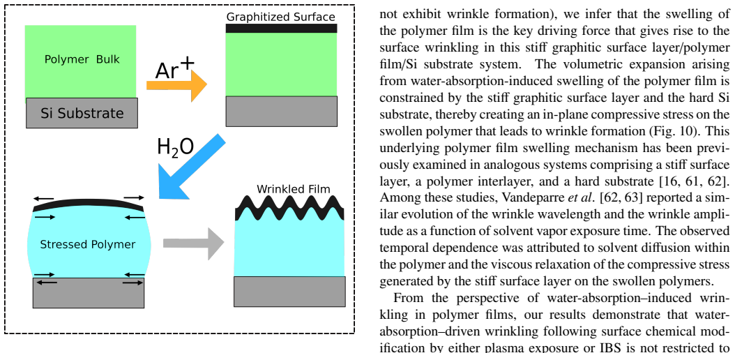
The load-bearing configuration is a three-layer stack: a stiff graphitized carbon skin formed by ion bombardment and estimated to be thinner than 10 nm, a water-absorbing polymer interlayer, and a rigid silicon substrate. The argument runs on the mismatch in swelling: water vapor penetrates the graphitic skin and hydrates the hydrophilic polymer, whose volumetric expansion is blocked by the skin and the substrate, building compressive in-plane stress that the surface relieves by buckling into wrinkles. The time-dependent growth of wrinkle wavelength and amplitude is read as evidence that swelling, not the ion beam itself, supplies the stress.
What would settle it
Measure the thickness or water mass of ion-bombarded p4VP films in a 60% RH atmosphere; if they wrinkle without gaining bulk water or swelling, the constrained-swelling mechanism cannot be the driver for p4VP. Conversely, if an ion-bombarded hydrophilic polymer that demonstrably swells in humid air never wrinkles even under a graphitic skin, the stiff-skin constraint story would need revision.
Extended reading notes
Core claim
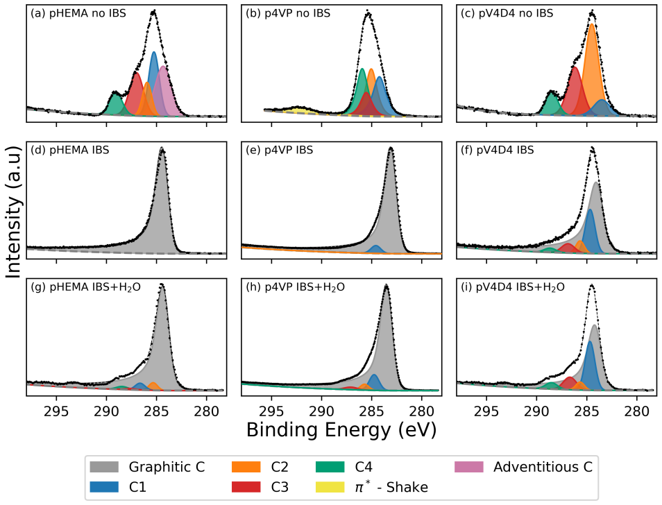
After 2 keV Ar+ bombardment, X-ray photoelectron spectroscopy shows that the surfaces of all three polymers are converted within the first minutes of sputtering into a graphitized carbon layer, roughly the top 10 nm, in which oxygen and nitrogen are largely stripped from the original polymer chemistry. For pHEMA and p4VP, exposure to about 60% relative humidity following bombardment then produces isotropic surface wrinkles whose wavelength and amplitude grow over days, while identical samples kept in dry argon stay flat. FTIR of pHEMA shows water-related hydroxyl and hydrogen-bonding signatures in the film bulk after humid exposure, and XPS shows water-induced oxidized carbon and nitrogen species at the surface, indicating water enters both the skin and the underlying polymer. The paper proposes that wrinkling is driven by water-absorption-induced swelling of the underlying polymer film, mechanically constrained by the stiff graphitic skin above and the rigid silicon substrate below. In pV4D4 the same ion treatment graphitizes the surface, but the polymer's low water uptake produces no appreciable swelling and therefore no wrinkles.
Load-bearing premise
For p4VP, the load-bearing premise is that it absorbs water throughout its bulk and swells after ion bombardment; the paper assumes this from surface XPS and published reports rather than measuring bulk swelling directly.
Editorial extensions
If this is right
- For any polymer film studied under ion bombardment, relative humidity after the run becomes a first-order variable: nominally identical ion doses can produce flat or wrinkled surfaces depending on whether humid air follows.
- Wrinkle size and pattern can be tuned by humidity-exposure time, since wavelength and amplitude grow over days in pHEMA and p4VP.
- Polymer hydrophilicity predicts which materials will wrinkle under this two-step treatment, with hydrophobic polymers acting as flat controls.
- The two-step route, ion beam to make a stiff skin and then water vapor to swell the bulk, extends water-vapor-driven wrinkling beyond plasma-treated PDMS to other polymer chemistries.
- Future reports of ion-beam-induced wrinkling must state the post-irradiation humidity history, or their morphology claims will be ambiguous.
Reading between the lines
- If the constrained-swelling picture is right, wrinkles should be patternable by masking the ion beam or by locally altering skin permeability, since the skin's water entry points would control where swelling can occur.
- A direct measurement of film thickness or water mass during humid exposure would connect the proposed swelling strain to the observed wrinkle growth; such a measurement would also give a quantitative stress estimate for the buckling condition.
- The comparison with pV4D4 suggests a practical test on the same polymer: tune water uptake chemically by copolymerizing pHEMA with a hydrophobic monomer and see whether wrinkle amplitude scales with absorbed water fraction.
- The graphitic skin is implicitly water-permeable; measuring water transport through an ion-beam-graphitized carbon layer would be a direct consequence of the mechanism.
Editorial analysis
A structured set of objections, weighed in public.
Referee Report
Summary. This manuscript reports an experimental study of ion beam sputtering (IBS) and humidity effects on three iCVD-grown polymer thin films: pHEMA, p4VP, and pV4D4. The central observation is that pHEMA and p4VP films on Si wrinkle only when IBS treatment is followed by exposure to water vapor at approximately 60% RH; films stored in a dry atmosphere after IBS remain flat, unbombarded films exposed to humidity remain flat, and pV4D4 remains flat under all tested conditions. AFM-based roughness and PSD analysis document the time evolution of wrinkle wavelength and amplitude. Ex situ and in situ XPS show that IBS converts the near-surface region into graphitic carbon within roughly two minutes of sputtering. ATR-FTIR measurements of the pHEMA film show water uptake in the bulk after humidity exposure. The authors propose that wrinkling results from water-absorption-induced swelling of the polymer film constrained by the stiff graphitic surface layer and the Si substrate, and attribute the absence of wrinkling in pV4D4 to its low water uptake. The manuscript claims the same bulk-water-absorption evidence for p4VP, but the FTIR measurements shown are for pHEMA only.
Significance. The paper's experimental core is solid and clearly presented: the two-by-two control structure (IBS vs. no IBS, humid vs. dry post-treatment), the in situ XPS experiments that rule out ambient oxidation as the source of the graphitization, and the quantitative AFM/PSD analysis with documented uncertainty propagation are all strengths. If the proposed mechanism holds, the results generalize wrinkling by constrained swelling beyond PDMS to hydrophilic polymer films, with a practical warning that ambient humidity must be controlled when interpreting IBS-induced polymer morphology. The main gap is evidentiary: bulk water uptake is demonstrated only for pHEMA, while the title-level claim covers both pHEMA and p4VP. Because the p4VP half of the mechanism rests on literature analogy and surface XPS rather than on a bulk measurement, the manuscript overreaches in its abstract and conclusion. This is fixable with additional data or with a carefully qualified claim.
major comments (3)
- [Abstract; Section 3.3; Section 5 (Conclusion)] The abstract and conclusion state that 'XPS and FTIR indicate water absorption in both the surface and the bulk of the films' for the wrinkling polymers pHEMA and p4VP, but the only bulk-sensitive FTIR data are presented for pHEMA. Section 3.3 is titled 'FTIR Study of Bulk Film Chemistry for pHEMA' and Figure 7 shows pHEMA spectra only; no FTIR spectra or band assignments are shown for p4VP (or pV4D4). The bulk swelling of p4VP is therefore inferred, not demonstrated, and the abstract overstates the evidence. Please either add bulk-sensitive measurements (e.g., ATR-FTIR or spectroscopic ellipsometry at controlled RH) for p4VP or explicitly restrict the bulk-absorption claim to pHEMA and rephrase the p4VP mechanism as conditional.
- [Section 4 (Discussion); Table 1; reference [59]] The p4VP half of the proposed mechanism relies on the claim that unmodified p4VP absorbs water and swells under the conditions of the experiment. The support offered is reference [59], a study of p(4-VP)-based materials for aqueous contaminant removal, which concerns materials in liquid water rather than a ~250-300 nm p4VP film at 60% RH, and the XPS data, which probe less than 10 nm. Neither establishes bulk swelling. In addition, Table 1 reports a large and unexplained change in the p4VP surface composition after humidity (N: 3% to 37%, C: 91% to 27%); this indicates a substantial surface chemical transformation that is not interpreted and does not, by itself, provide evidence for bulk water uptake. If p4VP does not swell in the bulk, the constrained-swelling mechanism cannot explain the observed p4VP wrinkling, so this point is load-bearing for half of the central claim.
- [Section 4; Figure 10] The proposed mechanism requires the IBS-modified surface layer to be mechanically stiff, but no mechanical measurement of the graphitized layer is presented; its thickness (<10 nm) and modulus are inferred from XPS chemistry and from the ion penetration depth. The wrinkling observations themselves imply a modulus mismatch, but the specific assignment of the skin as the stiff 'graphitic' phase is an assumption. A modulus estimate (e.g., nanoindentation or a buckling-based wrinkling analysis using the measured wavelength and film thickness) would materially strengthen the mechanism; without it, the discussion remains a plausible conjecture rather than a tested mechanical model.
minor comments (4)
- [Figure 2] The rendered labels in Figure 2 contain placeholder sequences such as '/uni00000053/uni0000002b/uni00000028/...' in place of readable chemical formulas; please regenerate the figure with proper text rendering.
- [Appendix A (captions of Figs. A.12 and A.13)] The phrase 'along the the white dash line' should read 'along the white dashed line' in the captions of Figures A.12 and A.13.
- [Equation (B.1)] Equation (B.1) and the following definition of the coefficients α′ and β′ appear garbled in the typeset text ('α=a S− as−α 1+4(...)'); the functional form is not readable and should be corrected.
- [Reference [10]] Reference [10] lacks the year of publication; the DOI resolves to a 2004 article and should be completed.
Circularity Check
No circularity: the wrinkling mechanism is assembled from external literature and independent controls; no fitted parameter is renamed as a prediction and no self-citation is load-bearing.
full rationale
The paper reports an empirical correlation (wrinkling occurs only after ion bombardment plus humidity, and pV4D4 does not wrinkle under the same conditions) and proposes a mechanism: water uptake swells the underlying polymer film, and that swelling is constrained by the stiff graphitized surface layer and the Si substrate. Each ingredient of the mechanism is supported by external references: graphitization of ion-bombarded polymers [21-23,47-51], swelling of hydrophilic polymers [58,59], low water absorption of siloxane polymers [60], and previous stiff-skin/polymer-interlayer wrinkling systems [16,61-63]. The only direct bulk water-uptake measurement (ATR-FTIR, Section 3.3, explicitly titled 'FTIR Study of Bulk Film Chemistry for pHEMA') is for pHEMA; for p4VP the bulk-swelling premise is inferred from the literature and from surface XPS changes. That is an evidence gap, not circularity: the paper does not fit a parameter to the wrinkling data and then 'predict' the wrinkling from that same fit, nor does it rely on a self-citation chain to exclude alternatives. The experimental controls (dry atmosphere, no IBS, and pV4D4 under identical conditions) provide independent, non-circular support for the claim that both ion bombardment and subsequent humidity are necessary for wrinkling in this system.
Assumptions & free parameters
assumptions (5)
- domain assumption The IBS-generated surface layer is a stiff, coherent graphitic skin with thickness below 10 nm.
- domain assumption Water absorption by pHEMA and p4VP causes volumetric swelling of the underlying film.
- domain assumption Water vapor can diffuse through the graphitized surface layer into the bulk polymer.
- domain assumption The silicon substrate is rigid and constrains out-of-plane expansion, converting swelling into in-plane compression.
- domain assumption pV4D4 has low water absorption and negligible swelling.
Cite this review
Pith. "Pith review of Wrinkling in Selected Polymer Thin Films Induced by Combined Ion Beam and Humidity Exposure." pith.science (2026). https://pith.science/paper/WPS7YT75
@misc{pith2026260809041,
  author       = {Pith},
  title        = {Pith review of: Wrinkling in Selected Polymer Thin Films Induced by Combined Ion Beam and Humidity Exposure},
  year         = {2026},
  howpublished = {\url{https://pith.science/paper/WPS7YT75}},
  note         = {Machine review of arXiv:2608.09041}
}
read the original abstract
This study investigates ion beam sputtering (IBS)-induced surface wrinkling phenomena in three polymers with varying hydrophilicity: poly-hydroxy-ethyl-methacrylate (pHEMA), poly-4-vinyl pyridine (p4VP), and poly-2,4,6,8-tetramethyl-2,4,6,8-tetravinylcyclotetrasiloxane (pV4D4). It is observed that pHEMA and p4VP films wrinkle only when exposed to ion bombardment and subsequent water vapor exposure. No wrinkling is observed in pV4D4 under these same conditions. X-ray photoelectron spectroscopy (XPS) and Fourier transform infrared spectroscopy (FTIR) are performed before IBS, after IBS, and after exposure to humidity. XPS shows that IBS drives chemical changes within the surface layer, creating a graphitized film at the surface. For the polymer films that exhibit wrinkling (pHEMA and p4VP), XPS and FTIR indicate water absorption in both the surface and the bulk of the films, resulting in swelling. We conjecture that the formation of wrinkles arises from this swelling being mechanically constrained by the rigid underlying silicon substrate and the stiff graphitized surface layer. In contrast, the absence of wrinkle formation in pV4D4 under the same experimental conditions can be attributed to its comparatively low water absorption and the correspondingly limited swelling response.
Figures


Figures from the paper (7 more)



Reference graph
Works this paper leans on
-
[59]
N. Sahiner, O. Ozay, N. Aktas, Aromatic organic con- taminant removal from an aqueous environment by p(4- VP)-based materials, Chemosphere 85 (5) 832–838. doi:10.1016/j.chemosphere.2011.06.087
-
[1]
N. Liu, Y . Lu, Z. Li, H. Zhao, Q. Yu, Y . Huang, J. Yang, L. Huang, Smart Wrinkled Interfaces: Patterning, Mor- phing, and Coding of Polymer Surfaces by Dynamic Anisotropic Wrinkling, Langmuir 40 (36) (2024) 18837– 18856. doi:10.1021/acs.langmuir.4c02162
-
[2]
R. N. Enright, L. C. Bradley, Self-Wrinkling Vapor- Deposited Polymer Films with Tunable Patterns, Ad- vanced Functional Materials 32 (39) (2022) 2204887. doi:10.1002/adfm.202204887
-
[3]
C.-M. Chen, S. Yang, Wrinkling instabilities in polymer films and their applications, Polymer International 61 (7) (2012) 1041–1047. doi:10.1002/pi.4223
-
[4]
T. R. Hendricks, I. Lee, Wrinkle-Free Nanome- chanical Film: Control and Prevention of Polymer Film Buckling, Nano Letters 7 (2) (2007) 372–379. doi:10.1021/nl062544q
-
[5]
E. Cerda, L. Mahadevan, Geometry and physics of wrinkling, Physical Review Letters 90 (2003) 074302. doi:10.1103/PhysRevLett.90.074302
-
[6]
Y .-C. Chen, A. J. Crosby, Wrinkling of inhomogeneously strained thin polymer films, Soft Matter 9 (1) (2013) 43–
work page 2013
-
[7]
J. S. Sharp, K. R. Thomas, M. P. Weir, Mechani- cally driven wrinkling instability in thin film polymer bilayers, Physical Review E 75 (1) (2007) 011601. doi:10.1103/PhysRevE.75.011601
Show all 73 references
-
[8]
Bowden, S
N. Bowden, S. Brittain, A. G. Evans, J. W. Hutchin- son, G. M. Whitesides, Spontaneous formation of or- dered structures in thin films of metals supported on an elastomeric polymer, Nature 393 (1998) 146–149. doi:10.1038/30193
1998 doi
-
[9]
A. Das, A. Banerji, R. Mukherjee, Programming fea- ture size in the thermal wrinkling of metal polymer bi- layer by modulating substrate viscoelasticity, ACS Ap- plied Materials & Interfaces 9 (2017) 23255–23262. doi:10.1021/acsami.7b08333
2017 doi
-
[10]
Okayasu, H
T. Okayasu, H. Zhang, D. G. Bucknall, G. A. D. Briggs, Spontaneous Formation of Ordered Lateral Pat- terns in Polymer Thin-Film Structures, Adv Funct Mate- rials 14 (11) 1081–1088. doi:10.1002/adfm.200305014
-
[11]
M.-W. Moon, S. H. Lee, J.-Y . Sun, K. H. Oh, A. Vaziri, J. W. Hutchinson, Wrinkled hard skins on polymers created by focused ion beam, Proceedings of the Na- tional Academy of Sciences 104 (4) (2007) 1130–1133. doi:10.1073/pnas.0610654104
2007 doi
-
[12]
M.-W. Moon, S. H. Lee, J.-Y . Sun, K. H. Oh, A. Vaziri, J. W. Hutchinson, Controlled formation of nanoscale wrinkling patterns on polymers using focused ion beam, Scripta Materialia 57 (8) (2007) 747–750. doi:10.1016/j.scriptamat.2007.06.043
2007 doi
-
[13]
M. W. Moon, E. K. Her, K. H. Oh, K. R. Lee, A. Vaziri, Sculpting on polymers using focused ion beam, Surface and Coatings Technology 202 (22) (2008) 5319–5324. doi:10.1016/j.surfcoat.2008.06.059
2008 doi
-
[14]
Jeong, H.-G
H.-C. Jeong, H.-G. Park, J. H. Lee, D.-S. Seo, Local- ized ion-beam irradiation-induced wrinkle patterns, ACS Applied Materials & Interfaces 7 (2015) 23216–23222. doi:10.1021/acsami.5b07147
2015 doi
-
[15]
Jeong, H.-G
H.-C. Jeong, H.-G. Park, J. H. Lee, Y . H. Jung, S. B. Jang, D.-S. Seo, Homogeneous self-aligned liquid crystals 13 on wrinkled-wall poly(dimethylsiloxane) via localised ion-beam irradiation, Scientific Reports 5 (2015) 8641. doi:10.1038/srep08641
2015 doi
-
[16]
Jeong, H.-G
H.-C. Jeong, H.-G. Park, Y . H. Jung, J. H. Lee, B.-Y . Oh, D.-S. Seo, Tailoring the Orientation and Periodicity of Wrinkles Using Ion-Beam Bom- bardment, Langmuir 32 (28) (2016) 7138–7143. doi:10.1021/acs.langmuir.6b01473
2016 doi
-
[17]
S. L. Arias, J. Devorkin, A. Civantos, J. P. Al- lain, Escherichia coli adhesion and biofilm for- mation on polydimethylsiloxane are independent of substrate stiffness, Langmuir 37 (2021) 16–25. doi:10.1021/acs.langmuir.0c00130
2021 doi
-
[18]
Breid, A
D. Breid, A. J. Crosby, Effect of stress state on wrinkle morphology, Soft Matter 7 (2011) 4490. doi:10.1039/c1sm05152k
2011 doi
-
[19]
D. B. H. Chua, H. T. Ng, S. F. Y . Li, Sponta- neous formation of complex and ordered structures on oxygen-plasma-treated elastomeric polydimethylsilox- ane, Applied Physics Letters 76 (2000) 721–723. doi:10.1063/1.125873
2000 doi
-
[20]
Ahmad, G
Z. Ahmad, G. Tyagi, S. Xiang, J. Heng, P. Patrício, P. I. C. Teixeira, C. M. Stafford, J. F. Douglas, J. T. Cabral, Surface Wrinkling of Plasma-Exposed PDMS is Caused by Water Vapor Sorption: An Optical En- vironmental Sensor, Adv Functional Materials 35 (47) 2509167. doi:10.1...
-
[21]
Calcagno, G
L. Calcagno, G. Compagnini, G. Foti, Structural modifi- cation of polymer films by ion irradiation, Nuclear Instru- ments and Methods in Physics Research Section B: Beam Interactions with Materials and Atoms 65 (1) (1992) IN7–
1992
-
[22]
C. M. Goodwin, Z. E. V oras, X. Tong, T. P. Beebe, Soft Ion Sputtering of PAni Studied by XPS, AFM, TOF-SIMS, and STS, Coatings 10 (10) (2020) 967. doi:10.3390/coatings10100967
2020 doi
-
[23]
S. N. Nesov, P. M. Korusenko, V . V . Bolotov, S. N. Povoroznyuk, D. A. Smirnov, Electronic struc- ture of nitrogen-containing carbon nanotubes irradi- ated with argon ions: XPS and XANES studies, Physics of the Solid State 59 (10) (2017) 2030–2035. doi:10.1134/S1063783417100286
2017 doi
-
[24]
Ne ˇcas, P
D. Ne ˇcas, P. Klapetek, Gwyddion: An open-source soft- ware for SPM data analysis, Open Physics 10 (1) 181–
-
[25]
S. R. Kane, P. D. Ashby, L. A. Pruitt, ATR-FTIR as a thickness measurement technique for hydrated polymer- on-polymer coatings, Journal of Biomedical Materials Re- search Part B: Applied Biomaterials 91B (2) (2009) 613–
2009
-
[26]
L. J. Fina, Depth Profiling of Polymer Surfaces with FT- IR Spectroscopy, Applied Spectroscopy Reviews 29 (3-4) (1994) 309–365. doi:10.1080/05704929408000562
1994 doi
-
[27]
H. Kaur, B. Rana, D. Tomar, S. Kaur, K. C. Jena, Fun- damentals of ATR-FTIR Spectroscopy and Its Role for Probing In-Situ Molecular-Level Interactions, in: D. K. Singh, M. Pradhan, A. Materny (Eds.), Modern Tech- niques of Spectroscopy, V ol. 13, Springer Singapore, Sin- gapor...
2021 doi
-
[28]
A. Götz, R. Nikzad-Langerodi, Y . Staedler, A. Bel- laire, J. Saukel, Apparent penetration depth in atten- uated total reflection Fourier-transform infrared (ATR- FTIR) spectroscopy ofAllium cepaL. epidermis and cuticle, Spectrochimica Acta Part A: Molecular and Biomolecular S...
2020
-
[29]
Laroche, J
G. Laroche, J. Fitremann, N. Gherardi, FTIR-ATR spec- troscopy in thin film studies: The importance of sampling depth and deposition substrate, Applied Surface Science 273 (2013) 632–637. doi:10.1016/j.apsusc.2013.02.095
2013 doi
-
[30]
Tougaard, Practical guide to the use of backgrounds in quantitative XPS, Journal of Vacuum Science & Technol- ogy A 39 (1) (2020) 011201
S. Tougaard, Practical guide to the use of backgrounds in quantitative XPS, Journal of Vacuum Science & Technol- ogy A 39 (1) (2020) 011201. doi:10.1116/6.0000661
2020 doi
-
[31]
D. G. Castner, B. D. Ratner, A. Hirao, S. Nakahama, Characterization of Poly(2-Hydroxyethyl Methacrylate) (PHEMA) by XPS, Surface Science Spectra 4 (1) (1996) 14–20. doi:10.1116/1.1247807
1996 doi
-
[32]
Estrade-Szwarckopf, XPS photoemission in carbona- ceous materials: A “defect” peak beside the graphitic asymmetric peak, Carbon 42 (8) (2004) 1713–1721
H. Estrade-Szwarckopf, XPS photoemission in carbona- ceous materials: A “defect” peak beside the graphitic asymmetric peak, Carbon 42 (8) (2004) 1713–1721. doi:10.1016/j.carbon.2004.03.005
2004 doi
-
[33]
Blume, D
R. Blume, D. Rosenthal, J.-P. Tessonnier, H. Li, A. Knop- Gericke, R. Schlögl, Characterizing Graphitic Carbon with X-ray Photoelectron Spectroscopy: A Step-by-Step Approach, ChemCatChem 7 (18) (2015) 2871–2881. doi:10.1002/cctc.201500344
2015 doi
-
[34]
Al-Gaashani, A
R. Al-Gaashani, A. Najjar, Y . Zakaria, S. Man- sour, M. A. Atieh, XPS and structural studies of high quality graphene oxide and reduced graphene ox- ide prepared by different chemical oxidation methods, Ceramics International 45 (11) (2019) 14439–14448. doi:10.1016/j.ceramint...
2019 doi
-
[35]
V . P. Afanas’ev, G. S. Bocharov, A. V . Eletskii, L. G. Lobanova, K. I. Maslakov, S. V . Savilov, Comparative Investigation of XPS Spectra of Oxidated Carbon Nan- otubes and Graphene, Biophysica 3 (2) (2023) 307–317. doi:10.3390/biophysica3020020
2023 doi
-
[36]
X. Chen, X. Wang, D. Fang, A review on C1s XPS-spectra for some kinds of carbon materials, Fullerenes, Nanotubes 14 and Carbon Nanostructures 28 (12) (2020) 1048–1058. doi:10.1080/1536383X.2020.1794851
2020
-
[37]
D. R. Baer, M. T. Camci, D. J. H. Cant, S. A. Cham- bers, H. Cohen, P. Aydogan Gokturk, D. J. Morgan, A. Shchukarev, P. M. A. Sherwood, S. Suzer, S. Tougaard, J. F. Watts, What more can be done with XPS? Highly in- formative but underused approaches to XPS data collec- tion an...
2025 doi
-
[38]
M. Maaz, T. Elzein, D. Dragoe, A. Bejjani, N. Jar- roux, C. Poulard, N. Aubry-Barroca, B. Nsouli, P. Roger, Poly(4-vinylpyridine)-modified silica for effi- cient oil/water separation, Journal of Materials Science 54 (2) (2019) 1184–1196. doi:10.1007/s10853-018-2888- x
2019 doi
-
[39]
P. S. Bagus, C. J. Nelin, M. Sassi, D. Baranowski, M. A. Sharp, T. Autrey, Z. Dohnálek, Z. Novotny, The XPS of pyridine: A combined theoretical and experimental anal- ysis, The Journal of Chemical Physics 162 (8) (2025) 084111. doi:10.1063/5.0249190
2025 doi
-
[40]
M. K. Kuntumalla, M. Attrash, R. Akhvlediani, S. Michaelson, A. Hoffman, Nitrogen bonding, work function and thermal stability of nitrided graphite surface: An in situ XPS, UPS and HREELS study, Applied Surface Science 525 (2020) 146562. doi:10.1016/j.apsusc.2020.146562
2020
-
[41]
Kusunoki, M
I. Kusunoki, M. Sakai, Y . Igari, S. Ishidzuka, T. Takami, T. Takaoka, M. Nishitani-Gamo, T. Ando, XPS study of nitridation of diamond and graphite with a nitro- gen ion beam, Surface Science 492 (3) (2001) 315–328. doi:10.1016/S0039-6028(01)01430-3
2001 doi
-
[42]
Titantah, D
J. Titantah, D. Lamoen, Carbon and nitrogen 1s energy levels in amorphous carbon nitride sys- tems: XPS interpretation using first-principles, Dia- mond and Related Materials 16 (3) (2007) 581–588. doi:10.1016/j.diamond.2006.11.048
2007 doi
-
[43]
Kondo, S
T. Kondo, S. Casolo, T. Suzuki, T. Shikano, M. Saku- rai, Y . Harada, M. Saito, M. Oshima, M. I. Trioni, G. F. Tantardini, J. Nakamura, Atomic-scale characterization of nitrogen-doped graphite: Effects of dopant nitrogen on the local electronic structure of the surrounding car...
2012 doi
-
[44]
Kiuchi, T
H. Kiuchi, T. Kondo, M. Sakurai, D. Guo, J. Naka- mura, H. Niwa, J. Miyawaki, M. Kawai, M. Oshima, Y . Harada, Characterization of nitrogen species incorpo- rated into graphite using low energy nitrogen ion sputter- ing, Physical Chemistry Chemical Physics 18 (1) (2016) 458–46...
2016 doi
-
[45]
Hellgren, R
N. Hellgren, R. T. Haasch, S. Schmidt, L. Hult- man, I. Petrov, Interpretation of X-ray photoelec- tron spectra of carbon-nitride thin films: New in- sights from in situ XPS, Carbon 108 (2016) 242–252. doi:10.1016/j.carbon.2016.07.017
2016 doi
-
[46]
Kiuchi, R
H. Kiuchi, R. Shibuya, T. Kondo, J. Nakamura, H. Niwa, J. Miyawaki, M. Kawai, M. Oshima, Y . Harada, Lewis Basicity of Nitrogen-Doped Graphite Observed by CO2 Chemisorption, Nanoscale Research Letters 11 (1) (2016)
2016
-
[47]
doi:10.1039/C2SM26822A
-
[48]
J. J. Végh, D. Nest, D. B. Graves, R. Bruce, S. Engel- mann, T. Kwon, R. J. Phaneuf, G. S. Oehrlein, B. K. Long, C. G. Willson, Near-surface modification of polystyrene by Ar+: Molecular dynamics simulations and experimen- tal validation, Applied Physics Letters 91 (23) (2007)...
2007 doi
-
[49]
J. J. Végh, D. Nest, D. B. Graves, R. Bruce, S. Engel- mann, T. Kwon, R. J. Phaneuf, G. S. Oehrlein, B. K. Long, C. G. Willson, Molecular dynamics simulations of near-surface modification of polystyrene: Bombardment with Ar+and Ar+/radical chemistries, Journal of Applied Physi...
2008 doi
-
[50]
Gokan, S
H. Gokan, S. Esho, Y . Ohnishi, Dry Etch Resistance of Organic Materials, Journal of The Electrochemical Soci- ety 130 (1) (1983) 143–146. doi:10.1149/1.2119642
1983 doi
-
[51]
Zekonyte, V
J. Zekonyte, V . Zaporojtchenko, F. Faupel, Investiga- tion of the drastic change in the sputter rate of poly- mers at low ion fluence, Nuclear Instruments and Meth- ods in Physics Research Section B: Beam Interactions with Materials and Atoms 236 (1-4) (2005) 241–248. doi:10....
2005 doi
- [52]
-
[53]
N. C. Norman, Periodicity and the s- and p-Block El- ements, Oxford Chemistry Primers, Oxford University Press, Oxford, New York, 1997
1997
-
[54]
Weinhold, R
F. Weinhold, R. West, The Nature of the Silicon– Oxygen Bond, Organometallics 30 (21) (2011) 5815–
2011
-
[55]
Delcorte, P
A. Delcorte, P. Bertrand, B. J. Garrison, Collision cas- cade and sputtering process in a polymer, The Jour- nal of Physical Chemistry B 105 (2001) 9474–9486. doi:10.1021/jp011099e. 15
2001 doi
-
[56]
H. T. Evensen, H. Jiang, K. W. Gotrik, F. Denes, R. W. Carpick, Transformations in wrinkle patterns: Coopera- tion between nanoscale cross-linked surface layers and the submicrometer bulk in wafer-spun, plasma-treated polydimethylsiloxane, Nano Letters 9 (2009) 2884–2890. doi:...
2009 doi
-
[57]
Kanski, B
M. Kanski, B. J. Garrison, Z. Postawa, Effect of Oxy- gen Chemistry in Sputtering of Polymers, The Journal of Physical Chemistry Letters 7 (8) (2016) 1559–1562. doi:10.1021/acs.jpclett.6b00514
2016 doi
-
[58]
Gulsen, A
D. Gulsen, A. Chauhan, Effect of water content on transparency, swelling, lidocaine diffusion in p-hema gels, Journal of Membrane Science 269 (2006) 35–48. doi:10.1016/j.memsci.2005.06.024
2006 doi
-
[60]
Reeja-Jayan, N
B. Reeja-Jayan, N. Chen, J. Lau, J. A. Kattirtzi, P. Moni, A. Liu, I. G. Miller, R. Kayser, A. P. Willard, B. Dunn, K. K. Gleason, A group of cyclic siloxane and silazane polymer films as nanoscale electrolytes for microbat- tery architectures, Macromolecules 48 (2015) 5222–52...
2015 doi
-
[61]
J. Y . Chung, A. J. Nolte, C. M. Stafford, Diffusion- controlled, self-organized growth of symmetric wrin- kling patterns, Advanced Materials 21 (2009) 1358–1362. doi:10.1002/adma.200803209
2009 doi
-
[62]
Vandeparre, S
H. Vandeparre, S. Gabriele, F. Brau, C. Gay, K. K. Parker, P. Damman, Hierarchical wrinkling patterns, Soft Matter 6 (2010) 5751. doi:10.1039/c0sm00394h
2010 doi
-
[63]
D. C. Hyun, U. Jeong, Substrate thickness: An effec- tive control parameter for polymer thin film buckling on pdms substrates, Journal of Applied Polymer Science 112 (2009) 2683–2690. doi:10.1002/app.29824
2009 doi
-
[64]
G. H. Major, V . Fernandez, N. Fairley, M. R. Linford, A detailed view of the Gaussian–Lorentzian sum and prod- uct functions and their comparison with the V oigt func- tion, Surface and Interface Analysis 54 (3) (2022) 262–
2022
-
[65]
T. R. Gengenbach, G. H. Major, M. R. Linford, C. D. Eas- ton, Practical guides for x-ray photoelectron spectroscopy (XPS): Interpreting the carbon 1s spectrum, Journal of Vacuum Science & Technology A 39 (1) (2021) 013204. doi:10.1116/6.0000682
2021 doi
-
[66]
G. H. Major, N. Fairley, P. M. A. Sherwood, M. R. Linford, J. Terry, V . Fernandez, K. Artyushkova, Prac- tical guide for curve fitting in x-ray photoelectron spec- troscopy, Journal of Vacuum Science & Technology A 38 (6) (2020) 061203. doi:10.1116/6.0000377. 16
2020 doi
-
[69]
Vandeparre, P
H. Vandeparre, P. Damman, Wrinkling of stimulore- sponsive surfaces: Mechanical instability coupled to diffusion, Physical Review Letters 101 (2008) 124301. doi:10.1103/PhysRevLett.101.124301
2008 doi
-
[127]
doi:10.1186/s11671-016-1344-6
-
[188]
doi:10.2478/s11534-011-0096-2
-
[269]
doi:10.1002/sia.7050
-
[422]
doi:10.1016/0168-583X(92)95077-5
-
[620]
doi:10.1002/jbm.b.31436
-
[5824]
doi:10.1021/om200675d
Reviewed August 14, 2026 · model on record in the stance chip above.
Discussion (0). Continue with ORCID to comment.