Biological Sex Determination in Cadavers Using Deep Learning Algorithms from Computed Tomography Images of Pelvis and Skull
Pith reviewed 2026-06-26 10:37 UTC · model grok-4.3
The pith
Deep learning models determine biological sex from CT scans of pelvis and skull in cadavers at 95.65 percent patient-level accuracy.
A machine-rendered reading of the paper's core claim, the machinery that carries it, and where it could break.
Core claim
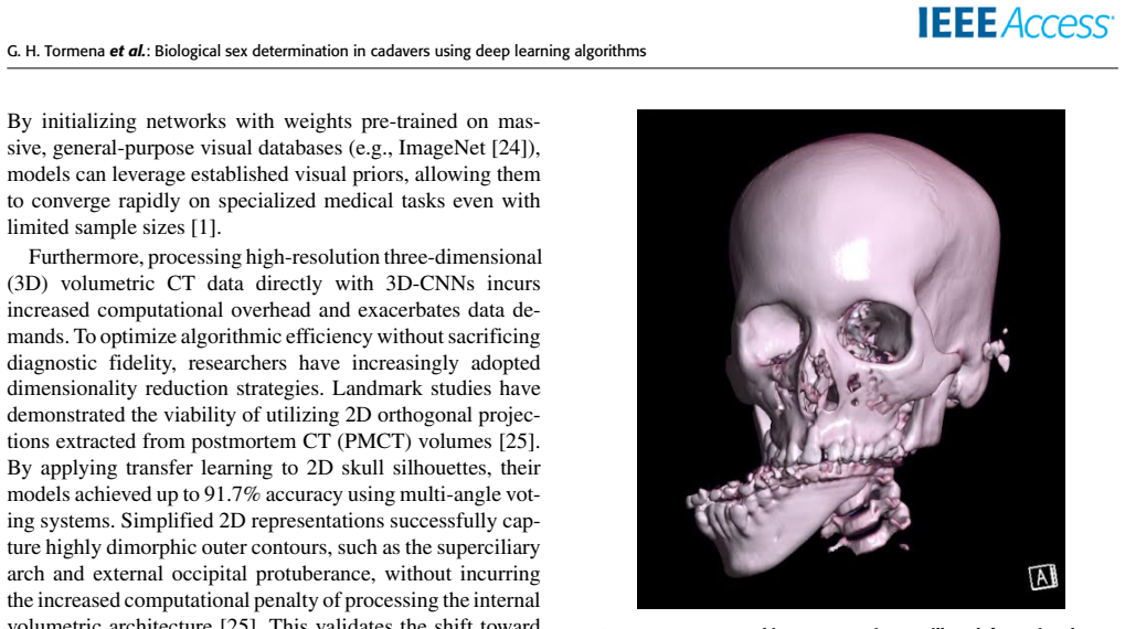
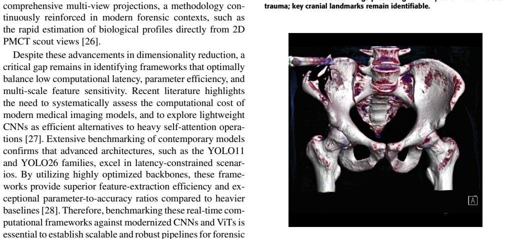
State-of-the-art deep learning models with transfer learning can classify biological sex from standardized 2D profile projections derived from 3D CT reconstructions of the pelvis and skull, achieving 95.65 percent patient-level accuracy, 92.86 percent recall, 94.36 percent F1-score, and 97.22 percent precision while remaining consistent across age ranges, preservation states, and trauma-damaged cases.
What carries the argument
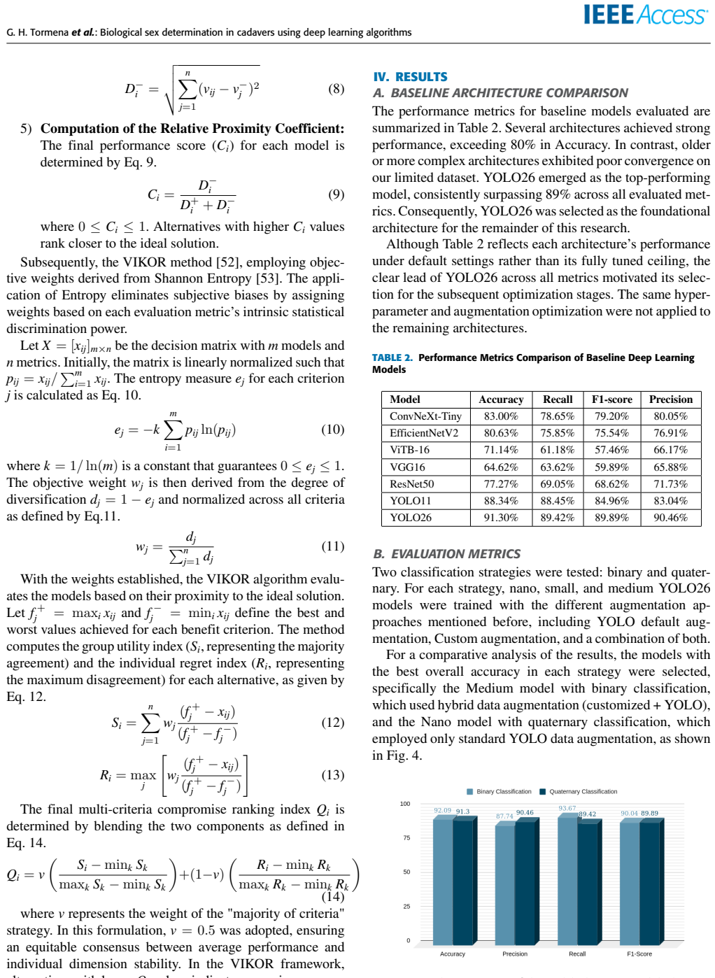
Transfer learning on deep networks (YOLO26, YOLO11, ConvNeXt-Tiny, EfficientNetV2, ViT-B16, VGG16, ResNet50) applied to 2D projections from 3D CT scans of pelvis and skull, with data augmentation to address limited samples.
If this is right
- Pelvis projections yield stronger and more consistent results than skull projections.
- Performance holds steady on cases that include trauma-related artifacts.
- Both binary sex classification and quaternary classification by sex plus anatomical region prove feasible.
- The method supplies an objective, high-speed alternative to manual skeletal analysis for forensic work.
Where Pith is reading between the lines
- The same projection-and-classification pipeline could be tested on other skeletal regions such as long bones for combined sex and age estimation.
- Integration into forensic imaging workstations might shorten turnaround time in mass-casualty or decomposed-remains scenarios.
- Retraining or domain adaptation would likely be needed before deployment on living patients or across diverse populations and scanner types.
Load-bearing premise
Data from a single forensic institute and one CT scanner captures enough variation to let the models work on scans from other centers and scanners.
What would settle it
Applying the trained models to CT scans collected at a second institution on a different scanner and obtaining patient-level accuracy below 80 percent.
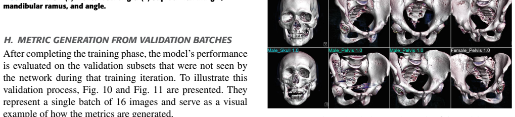
Figures
read the original abstract
Sexual identification of decomposed cadavers challenges traditional methods dependent on visual anthropological analysis. This study evaluates state-of-the-art deep learning (including YOLO26, YOLO11, ConvNeXt-Tiny, EfficientNetV2, ViT-B16, VGG16, and ResNet50) with transfer learning to automatically determine biological sex from forensic computed tomography (CT) scans. We analyzed 141 autopsied cadavers from the Forensic Medical Institute of Goi\^ania-GO, including a broad age range and varying conditions of preservation. The three-dimensional reconstructions of the pelvis and skull were converted into standardized two-dimensional profile projections, contributing to the study of this new technical approach. Data augmentation techniques compensated for sample limitations. Two scenarios were validated: binary and quaternary classification (one class per sex vs. one class per anatomical region of each sex). The best-performing model achieved highly consistent results on the pelvis region and still satisfactory performance on the skull region, reaching an overall patient-level accuracy of 95.65%, recall of 92.86%, F1- score of 94.36%, and precision of 97.22%, maintaining consistent performance across the evaluated cases, including those with trauma-related artifacts. Results indicate the technical feasibility of the methodology, demonstrating that deep learning models can provide objective, high-speed skeletal analysis. Since the study was conducted using data from a single institution and a single computed tomography scanner, further validation across multiple centers and scanners is required to assess the generalizability of the proposed approach
Editorial analysis
A structured set of objections, weighed in public.
Referee Report
Summary. The manuscript evaluates multiple deep learning architectures (YOLO26, YOLO11, ConvNeXt-Tiny, EfficientNetV2, ViT-B16, VGG16, ResNet50) with transfer learning on standardized 2D profile projections derived from 3D CT reconstructions of the pelvis and skull. Using a dataset of 141 autopsied cadavers from a single forensic institute, it compares binary and quaternary (sex × region) classification tasks, reports patient-level metrics up to 95.65% accuracy / 92.86% recall / 94.36% F1 / 97.22% precision (strongest on pelvis), and claims technical feasibility for objective, high-speed sex determination even in trauma cases, while noting the single-center/single-scanner limitation requires further multi-center validation.
Significance. If the reported performance generalizes, the work could supply a practical automated tool for forensic sex estimation in decomposed or fragmented remains where traditional visual methods are unreliable. The use of multiple modern architectures, data augmentation on a modest N, and explicit testing on trauma-affected cases are positive elements that support potential utility in high-throughput forensic settings.
major comments (2)
- [Abstract] Abstract: the headline performance figures (95.65% patient-level accuracy etc.) are presented without any description of train/test split strategy, cross-validation procedure, class-balance handling, or statistical significance testing. On a 141-case single-center corpus these omissions make it impossible to judge whether the metrics reflect genuine biological signal or overfitting to scanner-specific or demographic artifacts.
- [Abstract] Abstract: the claim that the method demonstrates 'technical feasibility' for objective skeletal analysis that 'works on trauma cases' rests on internal validation from one institution and one CT scanner. While the authors correctly flag the need for multi-center validation, this assumption is load-bearing for the generalization implied by the reported metrics and the quaternary classification / patient-level aggregation scheme; no quantitative evidence is supplied that learned features are driven by sex rather than acquisition parameters.
minor comments (1)
- [Abstract] Abstract: 'F1- score' contains an extraneous space before the hyphen.
Simulated Author's Rebuttal
We thank the referee for the constructive comments on our manuscript. We address each major comment point-by-point below, proposing revisions to the abstract where feasible while being transparent about limitations inherent to the single-center dataset.
read point-by-point responses
-
Referee: [Abstract] Abstract: the headline performance figures (95.65% patient-level accuracy etc.) are presented without any description of train/test split strategy, cross-validation procedure, class-balance handling, or statistical significance testing. On a 141-case single-center corpus these omissions make it impossible to judge whether the metrics reflect genuine biological signal or overfitting to scanner-specific or demographic artifacts.
Authors: The Methods section of the full manuscript describes the train/test split (held-out test set with patient-level aggregation), data augmentation to address class balance and sample size, and the use of transfer learning across the evaluated architectures. We agree that the abstract should briefly summarize these elements to allow independent assessment of the metrics. We will revise the abstract to include a concise statement on the validation strategy and augmentation. No formal statistical significance testing (e.g., p-values) was applied beyond standard performance metrics; this can be noted explicitly if required. revision: yes
-
Referee: [Abstract] Abstract: the claim that the method demonstrates 'technical feasibility' for objective skeletal analysis that 'works on trauma cases' rests on internal validation from one institution and one CT scanner. While the authors correctly flag the need for multi-center validation, this assumption is load-bearing for the generalization implied by the reported metrics and the quaternary classification / patient-level aggregation scheme; no quantitative evidence is supplied that learned features are driven by sex rather than acquisition parameters.
Authors: The manuscript already states the single-institution, single-scanner limitation and calls for multi-center validation. The standardized 2D projections and explicit inclusion of trauma-affected cases support feasibility claims within this cohort. However, we cannot supply quantitative evidence (e.g., via domain adaptation or multi-scanner ablation) that features are purely sex-driven versus acquisition artifacts, as no such external data exist in the study. We will revise the abstract to temper the feasibility language and reinforce the limitation. revision: partial
- Quantitative evidence that learned features are driven by biological sex rather than scanner-specific acquisition parameters, which would require multi-scanner datasets unavailable in the current single-center study.
Circularity Check
No circularity: purely empirical ML evaluation on held-out cadaver CT data
full rationale
The paper trains standard deep-learning classifiers (YOLO variants, ConvNeXt, EfficientNetV2, ViT, VGG16, ResNet50) with transfer learning on 141 single-center CT scans, converts 3-D reconstructions to 2-D projections, applies data augmentation, and reports patient-level accuracy/recall/F1/precision on internal test cases. No equations, first-principles derivations, fitted parameters renamed as predictions, or load-bearing self-citations appear in the derivation chain. Performance numbers are computed directly from model outputs versus ground-truth labels on the same dataset split; the single-institution limitation is explicitly flagged by the authors as requiring external validation rather than being smuggled in as a result. The evaluation is therefore self-contained against external benchmarks and contains no circular reduction.
Axiom & Free-Parameter Ledger
axioms (1)
- domain assumption Standard deep learning architectures with transfer learning can extract sex-discriminative features from 2D projections of CT bone images
Reference graph
Works this paper leans on
-
[1]
Zhang, ‘‘The application of forensic imaging to sex estimation: Focus on skull and pelvic structures,’’Perspectives in Legal and F orensic Sci- ences, vol
M. Zhang, ‘‘The application of forensic imaging to sex estimation: Focus on skull and pelvic structures,’’Perspectives in Legal and F orensic Sci- ences, vol. 1, 2024
2024
-
[2]
Krishan, P
K. Krishan, P . M. Chatterjee, T. Kanchan, S. Kaur, N. Baryah, and R. K. Singh, ‘‘A review of sex estimation techniques during examination of skeletal remains in forensic anthropology casework,’’F orensic Science International, vol. 261, pp. 165.e1–165.e8, apr 2016. [Online]. Available: https://www.sciencedirect.com/science/article/pii/S0379073816300202
2016
-
[3]
Seifert, L
Z. Seifert, L. Friedl, K. Chaumoitre, and J. Brůžek, ‘‘Applicability and lim- itations of sex assessment based on foramen magnum,’’F orensic Science International, vol. 271, pp. 126.e1–126.e9, feb 2017. [Online]. Available: https://www.sciencedirect.com/science/article/pii/S0379073816305308
2017
-
[4]
R. Lye, H. Min, J. Dowling, Z. Obertová, M. Estai, N. A. Bachtiar, and D. Franklin, ‘‘Deep learning versus human assessors: forensic sex estimation from three-dimensional computed tomography scans.’’ Scientific Reports, vol. 14, 2024. [Online]. Available: https://www.nature. com/articles/s41598-024-81718-y
2024
-
[5]
Tournois, V
L. Tournois, V . Trousset, D. Hatsch, T. Delabarde, B. Ludes, and T. Lefèvre, ‘‘Artificial intelligence in the practice of forensic medicine: a scoping review,’’International Journal of Legal Medicine, vol. 138, 12 2023
2023
-
[6]
Galante, R
N. Galante, R. Cotroneo, D. Furci, G. Lodetti, and M. B. Casali, ‘‘Ap- plications of artificial intelligence in forensic sciences: Current potential benefits, limitations and perspectives,’’International Journal of Legal Medicine, vol. 137, pp. 445–458, 2023
2023
-
[7]
Y . Cao, Y . Ma, X. Y ang, J. Xiong, Y . Wang, J. Zhang, Z. Qin, Y . Chen, D. N. Vieira, F. Chen, J. Zhang, and P . Huang, ‘‘Use of deep learning in forensic sex estimation of virtual pelvic models from the han population,’’ F orensic Sciences Research, vol. 7, no. 3, pp. 540–549, 07 2022. [Online]. Available: https://doi.org/10.1080/20961790.2021.2024369
-
[8]
Kourounis, A
G. Kourounis, A. A. Elmahmudi, B. Thomson, J. Hunter, H. Ugail, and C. Wilson, ‘‘Computer image analysis with artificial intelligence: a practi- cal introduction to convolutional neural networks for medical profession- als,’’Postgraduate Medical Journal, vol. 99, no. 1178, 2023
2023
-
[9]
De-Giorgio, G
F. De-Giorgio, G. Ciasca, G. Fecondo, A. Mazzini, R. Di Santo, M. De Spir- ito, and V . L. Pascali, ‘‘Post mortem computed tomography meets ra- diomics: a case series on fractal analysis of post mortem changes in the brain,’’International Journal of Legal Medicine, vol. 136, pp. 719–727, 2022
2022
-
[10]
E. Ortiz Rosa, E. M. Crosato, C. C. Castro, R. E. Oliveira, and M. G. H. Biazevic, ‘‘Comparative study of sex estimates in adult skulls using direct measurement and tomographic image reconstruction,’’ Brazilian Oral Research, vol. 37, 2023. [Online]. Available: https: //doi.org/10.1590/1807-3107bor-2023.vol37.0064
-
[11]
Guareschi, S
E. Guareschi, S. Poggesi, M. Palmesino, and P . Magni, ‘‘The presence of the human auditory ossicles—detected postmortem by ct scan—as a taphonomic indicator,’’F orensic Sciences, vol. 3, pp. 560–570, 11 2023
2023
-
[12]
C. Lin, A. P . Y oon, C.-W. Wang, T. Chao, K. C. Chung, and C.-F. Kuo, ‘‘Effects of image degradation on deep neural network classification of scaphoid fracture radiographs: Comparison study of different noise types,’’ JMIR Med Inform, vol. 14, p. e65596, Jan 2026
2026
-
[13]
R. Morán-Torres, K. Feld, J. Hesser, Y . M. Taalab, and K. Y en, ‘‘Artificial intelligence and computer vision in forensic sciences,’’ Rechtsmedizin, vol. 35, no. 4, pp. 219–225, 08 2025. [Online]. Available: https://doi.org/10.1007/s00194-025-00775-3
-
[14]
PL, ‘‘Sexing skulls using discriminant function analysis of visually assessed traits,’’American Journal of Physical Anthropology, vol
W. PL, ‘‘Sexing skulls using discriminant function analysis of visually assessed traits,’’American Journal of Physical Anthropology, vol. 136, no. 1, pp. 39–50, 05 2008. [Online]. Available: https://doi.org/10.1002/ ajpa.20776
2008
-
[15]
T. W. Phenice, ‘‘A newly developed visual method of sexing the os pubis,’’ American Journal of Physical Anthropology, vol. 30, no. 2, pp. 297–301, 1969
1969
-
[16]
X. Wang, G. Liu, Q. Wu, Y . Zheng, F. Song, and Y . Li, ‘‘Sex estimation techniques based on skulls in forensic anthropology: A scoping review,’’ PLoS ONE, vol. 19, no. 12, 12 2024. VOLUME 11, 2023 13 G. H. Tormenaet al.: Biological sex determination in cadavers using deep learning algorithms
2024
-
[17]
A. R. Klales, S. D. Ousley, and J. M. V ollner, ‘‘A revised method of sexing the human innominate using phenice’s nonmetric traits and statistical methods,’’American Journal of Physical Anthropology, vol. 149, no. 1, pp. 104–114, 2012
2012
-
[18]
P . Shah, Y . Kumar, and T. Bhowmik, ‘‘Deep learning techniques for the diagnosis and detection of orthopedic conditions: A systematic review of recent advances and challenges,’’Archives of Computational Methods in Engineering, vol. 33, 09 2025
2025
-
[19]
L. Chen, J. Chen, H. Hajimirsadeghi, and G. Mori, ‘‘Adapting grad-cam for embedding networks,’’ in2020 IEEE Winter Conference on Applications of Computer Vision (WACV), 2020, pp. 2783–2792
2020
-
[20]
Anees, R
W. Anees, R. Silva, A. Khan, J. Murray, L. Scavassini, M. Burle, N. Angelakopoulos, M. H. Napimoga, L. Porto, A. Abade, and A. Franco, ‘‘Maxillary sinus classification for sex and age using 23 artificial intelligence architectures,’’Scientific Reports, vol. 16, no. 1, p. 5716, 01
-
[21]
Available: https://doi.org/10.1038/s41598-026-36112-1
[Online]. Available: https://doi.org/10.1038/s41598-026-36112-1
-
[22]
A. Bulut, M. B. Aşkın, and G. Çınarer, ‘‘Comparative analysis of third molar segmentation performance between sexes using deep learning models,’’Diagnostics, vol. 16, no. 7, p. 977, 03 2026. [Online]. Available: https://doi.org/10.3390/diagnostics16070977
-
[23]
S. N. Batool, J. Y ang, G. Gilanie, A. Latif, S. Y asin, A. Ikram, and L. Y . Por, ‘‘Forensic radiology: A robust approach to biological profile estimation from bone image analysis using deep learning,’’Biomedical Signal Processing and Control, vol. 105, p. 107661, 2025. [Online]. Available: https://www.sciencedirect.com/science/article/pii/S1746809425001727
2025
-
[24]
K. Weiss, T. M. Khoshgoftaar, and D. Wang, ‘‘A survey of transfer learning,’’Journal of Big Data, vol. 3, no. 1, p. 9, 2016. [Online]. Available: https://doi.org/10.1186/s40537-016-0043-6
-
[25]
A. Krizhevsky, I. Sutskever, and G. E. Hinton, ‘‘Imagenet classification with deep convolutional neural networks,’’Commun. ACM, vol. 60, no. 6, p. 84–90, May 2017. [Online]. Available: https://doi.org/10.1145/3065386
-
[26]
T. Seo, Y . Y oon, Y . Kim, Y . Usumoto, N. Eto, Y . Sadamatsu, R. Tadakuma, and J. Morishita, ‘‘Sex estimation using skull silhouette images from postmortem computed tomography by deep learning,’’Scientific Reports, vol. 14, p. 22689, 09 2024
2024
-
[27]
Provost, M.-E
C. Provost, M.-E. Richard, T. Delabarde, G. Hmeydia, A. Berre, W. Hassen, O. Naggara, L. Hamza, B. Ludes, C. Oppenheim, and J. Benzakoun, ‘‘Age and sex estimation using post-mortem ct scout view,’’International Journal of Legal Medicine, pp. 1–14, 03 2026
2026
-
[28]
Z. Xue, P . Pi, Z. Liu, Z. Zeng, and Z. Sun, ‘‘Mednext for accurate medical image classification and segmentation: A lightweight transformer-style convolutional neural network,’’PLOS One, vol. 21, 01 2026
2026
-
[29]
Sapkota, R
R. Sapkota, R. H. Cheppally, A. Sharda, and M. Karkee, ‘‘Y olo26: Key architectural enhancements and performance benchmarking for real-time object detection,’’ 2026
2026
-
[30]
Mustra, K
M. Mustra, K. Delac, and M. Grgic, ‘‘Overview of the dicom standard,’’ in 2008 50th International Symposium ELMAR, vol. 1, 2008, pp. 39–44
2008
-
[31]
Levoy, ‘‘ Display of Surfaces from V olume Data ,’’IEEE Computer Graphics and Applications, vol
M. Levoy, ‘‘ Display of Surfaces from V olume Data ,’’IEEE Computer Graphics and Applications, vol. 8, no. 03, pp. 29–37, 1988. [Online]. Available: https://doi.ieeecomputersociety.org/10.1109/38.511
-
[32]
Mason, ‘‘pydicom/pydicom: pydicom 3.0.1,’’ 2024
D. Mason, ‘‘pydicom/pydicom: pydicom 3.0.1,’’ 2024. [Online]. Available: https://doi.org/10.5281/zenodo.13824606
-
[33]
Tan and Q
M. Tan and Q. Le, ‘‘Efficientnetv2: Smaller models and faster training,’’ in International Conference on Machine Learning (ICML). PMLR, 2021, pp. 10 096–10 106
2021
-
[34]
Z. Liu, H. Mao, C.-Y . Wu, C. Feichtenhofer, T. Darrell, and S. Xie, ‘‘A convnet for the 2020s,’’ pp. 11 976–11 986, 2022
2022
-
[35]
A. Dosovitskiy, L. Beyer, A. Kolesnikov, D. Weissenborn, X. Zhai, T. Un- terthiner, M. Dehghani, M. Minderer, G. Heigold, S. Gellyet al., ‘‘An image is worth 16x16 words: Transformers for image recognition at scale,’’ arXiv preprint arXiv:2010.11929, 2021
Pith/arXiv arXiv 2010
-
[36]
K. Simonyan and A. Zisserman, ‘‘V ery deep convolutional networks for large-scale image recognition,’’arXiv preprint arXiv:1409.1556, 2014
Pith/arXiv arXiv 2014
-
[37]
K. He, X. Zhang, S. Ren, and J. Sun, ‘‘Deep residual learning for image recognition,’’ inProceedings of the IEEE Conference on Computer Vision and Pattern Recognition (CVPR), 2016, pp. 770–778
2016
-
[38]
Jocher, J
G. Jocher, J. Qiu, and M. Najibi, ‘‘Ultralytics yolo11,’’ 2024. [Online]. Available: https://github.com/ultralytics/ultralytics
2024
-
[39]
Jocher, A
G. Jocher, A. Chaurasia, and J. Qiu, ‘‘Ultralytics yolov26,’’ 2026. [Online]. Available: https://github.com/ultralytics/ultralytics
2026
-
[40]
A. Wong, M. Famuori, M. J. Shafiee, F. Li, B. Chwyl, and J. Chung, ‘‘Y olo nano: a highly compact you only look once convolutional neural network for object detection,’’ 2019. [Online]. Available: https: //arxiv.org/abs/1910.01271
arXiv 2019
-
[41]
A. Buslaev, V . I. Iglovikov, E. Khvedchenya, A. Parinov, M. Druzhinin, and A. A. Kalinin, ‘‘Albumentations: Fast and flexible image augmentations,’’ Information, vol. 11, no. 2, p. 125, 2020. [Online]. Available: https: //doi.org/10.3390/info11020125
-
[42]
O’Gara and K
S. O’Gara and K. McGuinness, ‘‘Comparing data augmentation strategies for deep image classification,’’ inIMVIP 2019: Irish Machine Vision & Image Processing, Technological University Dublin, Dublin, Ireland, August 2019
2019
-
[43]
You Only Look Once: Unified, Real -Time Object Detection[J].IEEE, 2016.DOI:10.1109/CVPR.2016.91
J. Redmon, S. Divvala, R. Girshick, and A. Farhadi, ‘‘Y ou only look once: Unified, real-time object detection,’’ inProceedings of the IEEE Conference on Computer Vision and Pattern Recognition, 2016, pp. 779–788. [Online]. Available: https://doi.org/10.1109/CVPR.2016.91
-
[44]
YOLOv4: Optimal Speed and Accuracy of Object Detection
A. Bochkovskiy, C. Y . Wang, and H. Y . M. Liao, ‘‘Y olov4: Optimal speed and accuracy of object detection,’’arXiv preprint arXiv:2004.10934, 2020. [Online]. Available: https://doi.org/10.48550/arXiv.2004.10934
work page internal anchor Pith review Pith/arXiv arXiv doi:10.48550/arxiv.2004.10934 2004
-
[45]
Wong and P .-Y
T.-T. Wong and P .-Y . Y eh, ‘‘Reliable accuracy estimates from k-fold cross validation,’’IEEE Transactions on Knowledge and Data Engineering, vol. 32, no. 8, pp. 1586–1594, 2020
2020
-
[46]
Sokolova and G
M. Sokolova and G. Lapalme, ‘‘A systematic analysis of performance measures for classification tasks,’’Information Processing & Management, vol. 45, no. 4, pp. 427–437, 2009
2009
-
[47]
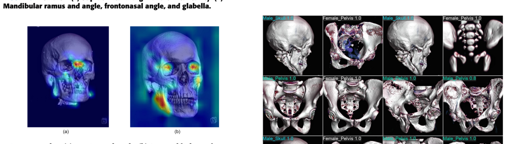
R. R. Selvaraju, M. Cogswell, A. Das, R. V edantam, D. Parikh, and D. Batra, ‘‘Grad-cam: Visual explanations from deep networks via gradient-based localization,’’International Journal of Computer Vision, vol. 128, no. 2, p. 336–359, Oct. 2019. [Online]. Available: http://dx.doi.org/10.1007/s11263-019-01228-7
-
[48]
K. Wang, S. Yin, Y . Wang, and S. Li, ‘‘Explainable deep learning for medical image segmentation with learnable class activation mapping,’’ in2023 2nd Asia Conference on Algorithms, Computing and Machine Learning (CACML 2023). ACM, 2023, pp. 210–215
2023
-
[49]
V enema, D
J. V enema, D. Peula, J. Irurita, and P . Mesejo, ‘‘Employing deep learning for sex estimation of adult individuals using 2d images of the humerus,’’ Neural Computing and Applications, vol. 35, no. 8, pp. 5987–5998, 2023
2023
-
[50]
Lambora, K
A. Lambora, K. Gupta, and K. Chopra, ‘‘Genetic algorithm- a literature review,’’ in2019 International Conference on Machine Learning, Big Data, Cloud and Parallel Computing (COMITCon), 2019, pp. 380–384
2019
-
[51]
J. M. Johnson and T. M. Khoshgoftaar, ‘‘Survey on deep learning with class imbalance,’’Journal of Big Data, vol. 6, no. 1, p. 27, 03 2019. [Online]. Available: https://doi.org/10.1186/s40537-019-0192-5
-
[52]
Çelikbilek & Fatih Tüysüz, ‘‘An in-depth review of theory of the topsis method: An experimental analysis,’’Journal of Management Analytics, 2020
Y . Çelikbilek & Fatih Tüysüz, ‘‘An in-depth review of theory of the topsis method: An experimental analysis,’’Journal of Management Analytics, 2020
2020
-
[53]
Mardani, E
A. Mardani, E. K. Zavadskas, K. Govindan, A. Amat Senin, and A. Jusoh, ‘‘Vikor technique: A systematic review of the state of the art literature on methodologies and applications,’’Sustainability, vol. 8, no. 1, 2016. [Online]. Available: https://www.mdpi.com/2071-1050/8/1/37
2016
-
[54]
Saraiva, ‘‘On shannon entropy and its applications,’’Kuwait Journal of Science, vol
P . Saraiva, ‘‘On shannon entropy and its applications,’’Kuwait Journal of Science, vol. 50, no. 3, p. 194–199, 2023
2023
-
[55]
S. S. Moosa, M. H. R. Shaikh, M. Khwaja, S. A. H. Shaikh, F. B. Siddiqui, S. R. H. Daimi, S. D. Hiware, E. E. Ismail, and Y . Begum, ‘‘Sexual di- morphic parameters of femur: A clinical guide in orthopedics and forensic studies,’’Journal of Medicine and Life, vol. 14, no. 6, pp. 762–768, 2021
2021
-
[56]
S. M. Mani, Y . S. Ahamed, P . Ambiga, V . Ramalingam, G. Sivaraman, and N. Balan, ‘‘Evaluation of orbital morphometry using 3d computed to- mographic images in biological sex determination: a retrospective study,’’ Journal of Indian Academy of Oral Medicine and Radiology, vol. 32, no. 4, p. 390, 2020
2020
-
[57]
E. D. Kalkan, A. Baransel, M. Akşamoğlu, M. Akbaba, and H. Kalkan, ‘‘Sex determination using craniometric parameters: A computed tomography- based assessment,’’Adli Tıp Dergisi, vol. 39, no. 2, p. 125–135, 2025. 14 VOLUME 11, 2023 G. H. Tormenaet al.: Biological sex determination in cadavers using deep learning algorithms GIOVANNA HERCULANO TORMENAis cur-...
2025
discussion (0)
Sign in with ORCID, Apple, or X to comment. Anyone can read and Pith papers without signing in.