REVIEW 4 major objections 5 minor 31 references
Real-Time Guidewire Tip Tracking Using a Siamese Network for Image-Guided Endovascular Procedures
T0 review · 4 major / 5 minor · reviewed 2026-08-06 · deepseek-v4-flash
Pith's one-line read A Siamese network with dual attention tracks guidewire tips in real-time DSA images with 0.421 mm mean error and transfers zero-shot to a robotic phantom.
desk verdict A sensible Siamese tracker for guidewire tips with a real robotic validation, but every mm accuracy number is unverifiable because the pixel-to-mm conversion is never reported. read the letter →
The pith
A machine-rendered reading of the paper's core claim, the machinery that carries it, and where it could break.
The reading
What carries the argument
The central mechanism is the Directional Edge-Enhanced Attention Network (DEAN) operating inside a Siamese tracker. DEAN computes directional edge maps with learnable Sobel-style kernels, applies channel attention that pools global context and spatial attention that fuses the original and edge-enhanced features, then multiplies both attention maps into the feature tensor. This selectively emphasizes the guidewire's thin, elongated structure and suppresses vascular background, artifacts, and noise. A transformer encoder provides self-attention across the template and search features, and a frequency-domain normalized cross-correlation yields a translation-equivariant response map that feeds classification, centerness, and regression heads; the multi-task loss combines GIoU, binary cross-entropy, and Smooth L1. Together these components convert the template-matching paradigm into a segmentation-free, real-time localization pipeline.
What would settle it
Take the three test DSA sequences, read the pixel spacing from their DICOM headers, and recompute center errors from the predicted and ground-truth box centers; if the spacing varies by sequence or is absent, the claimed $0.421$ mm mean cannot be reproduced. A second check is annotating a public DSA dataset with millimeter ground truth and running the released model; the method would be falsified if the observed mean error exceeds the reported value by more than the standard deviation.
Extended reading notes
Core claim
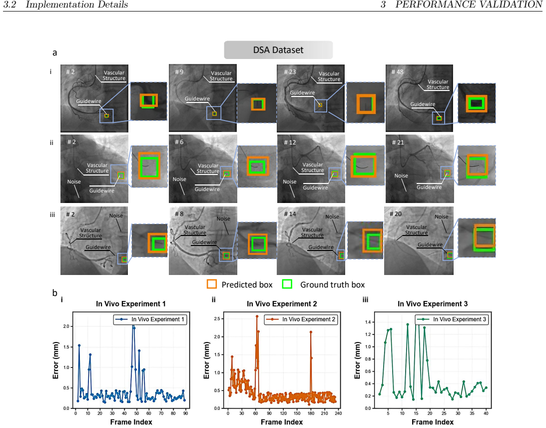
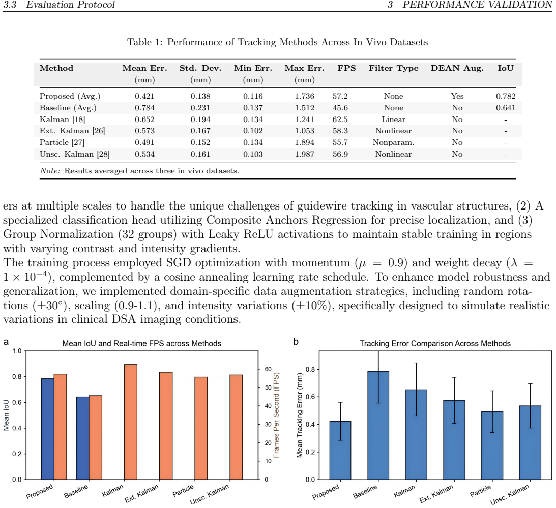
On its own terms, the paper's central claim is that a Siamese template-matching tracker, augmented by edge-enhanced dual attention, can localize the guidewire tip in intraoperative DSA images with sub-millimeter accuracy at clinical frame rates. The network combines self-attention through a transformer encoder with channel and spatial attention in DEAN, and uses frequency-domain cross-correlation between the template and search patches to produce a dense similarity map. Guided by separate classification, centerness, and regression heads trained with GIoU, BCE, and Smooth L1 losses, the model achieves a mean error of $0.421\pm 0.138$ mm and an IoU of $0.782$ on three held-out clinical sequences, outperforming Kalman, extended Kalman, particle, and unscented Kalman filters in mean error. The same weights, with no fine-tuning, track the tip in a robotic RGB phantom at 38 and 36 FPS, supporting the claim that the tracker can act as the perception input for autonomous endovascular navigation.
Load-bearing premise
Every millimeter-level accuracy figure assumes an unstated pixel-to-millimeter calibration of the DSA sequences, because ground-truth boxes are described as roughly 12×12 pixels while errors are reported in millimeters; if the pixel spacing is not fixed and known, none of the reported errors can be verified or compared across methods.
Editorial extensions
If this is right
- At 57.2 FPS, the tracker exceeds the 15–30 FPS range the paper cites as needed for smooth intraoperative visualization, so it can feed continuous tip coordinates during an intervention.
- Because tracking does not depend on a segmentation step, it avoids the failure mode the paper attributes to UNet-based endpoint localization when vessel boundaries are obscured by motion blur or contrast washout.
- The mean IoU of 0.782 versus 0.641 for the no-DEAN baseline implies the edge-enhanced attention module is what closes most of the accuracy gap, not just the Siamese backbone.
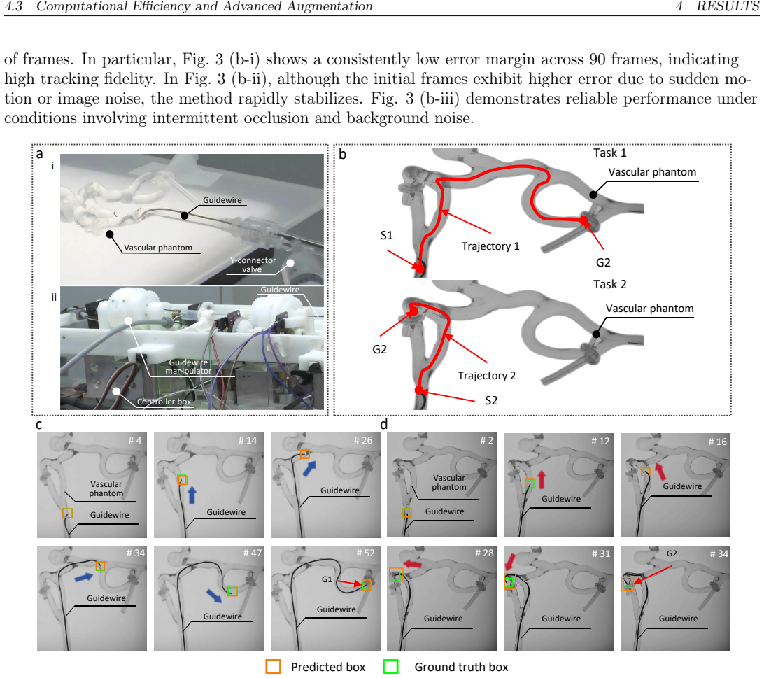
- Zero-shot transfer to the robotic RGB phantom with mean errors of 0.708 mm and 0.148 mm indicates the same model can serve as a perception module for autonomous guidewire navigation without retraining on the robotic camera.
- The maximum clinical error of 1.736 mm bounds the worst-case localization drift, suggesting the tracker remains clinically plausible even during transient artifacts.
Reading between the lines
- My inference: the large difference between the two robotic tasks (0.708 vs 0.148 mm) likely reflects imaging quality more than tracking difficulty, so the zero-shot numbers should be read as a range of domain-gap conditions rather than a single transfer accuracy.
- My inference: because the paper reports errors in millimeters while annotations are described as about 12×12 pixel boxes, publishing the DICOM pixel spacing for each sequence would let other groups reproduce the comparison; without it, the headline accuracy cannot be independently checked.
- My inference: the frame-to-frame template update may be the first failure point under prolonged occlusion; a natural extension is to add a re-detection or template-reinitialization mechanism and test recovery after the tip disappears behind a bifurcation.
- My inference: the same Siamese-plus-edge-attention design should transfer to other thin instruments such as catheters, needles, or guidewire shafts, where the same linear-edge prior applies; a cheap test is to train on DSA frames with the shaft tip annotated instead of the distal tip.
Editorial analysis
A structured set of objections, weighed in public.
Referee Report
Summary. The paper proposes a Siamese-network tracker with self- and cross-attention mechanisms and a Directional Edge-Enhanced Attention Network (DEAN) for real-time guidewire tip tracking in DSA images. The method is validated on three clinical DSA sequences, reporting a mean localization error of 0.421 +/- 0.138 mm, a maximum error of 1.736 mm, a mean IoU of 0.782, and 57.2 FPS. It further reports zero-shot transfer to a robotic vascular phantom with errors of 0.708 +/- 0.695 mm and 0.148 +/- 0.057 mm in two navigation tasks. The central claim is that the framework is a real-time, segmentation-free perception module for image-guided endovascular procedures and robot-assisted navigation.
Significance. If the reported figures are reproducible, the contribution is practically useful: a real-time guidewire-tip tracker that avoids a segmentation dependency, with evidence from clinical DSA sequences and a robotic phantom. The architecture is described in enough detail to be reimplemented, and the inclusion of a DEAN ablation baseline is a useful check. The paper also acknowledges important limitations, including the small proprietary dataset and the clinical-to-robotic domain gap. However, the central quantitative claims are currently not fully verifiable because the pixel-to-millimeter conversion is not reported and because the robotic ground-truth reference standard is described inconsistently. These issues are local and fixable, but they must be resolved before the accuracy figures can be accepted.
major comments (4)
- [Section 3.1, Section 3.3, Table 1] The reported millimeter accuracies are not reproducible because no pixel-to-millimeter conversion is provided. Section 3.1 states that ground-truth boxes average 12x12 pixels with a standard deviation of +/-3 pixels, while Section 3.3 and Table 1 report center error in millimeters; no DICOM pixel spacing, detector calibration factor, or mm-per-pixel value is given for any of the three DSA sequences. If the sequences come from different patients or procedures, their magnification and pixel spacing may differ, so even a single scale factor may not be well defined. This omission underpins every error value in the paper, including the 0.421 mm mean, the 1.736 mm maximum, the 0.708/0.148 mm robotic errors, and the 'sub-pixel accuracy' claim in Section 6. Please report the per-sequence pixel spacing or another explicit, verifiable conversion and discuss whether a single conversion factor is valid across all three sequences.
- [Section 3.1, Section 4.4, Figure 5] The reference standard for the robotic experiments is internally inconsistent. Section 3.1 says that the robot's registration system provides ground truth with +/-0.1 mm accuracy, but Section 4.4 and the Figure 5 captions describe the orange boxes as manually labeled or manually annotated ground truth. The claimed 0.708 and 0.148 mm errors therefore have an unclear reference: if the ground truth is manual labels in RGB images, the errors are not measured against the robot-registration ground truth, and the +/-0.1 mm accuracy statement is irrelevant. Please state which reference standard was used for each robotic task and explain how the manual annotations relate to the robot registration system.
- [Section 4.1, Table 1] The comparison is not sufficient to support the claim that the method outperforms existing approaches. The baselines in Table 1 are Kalman, extended Kalman, particle, and unscented Kalman filters, plus a single 'Baseline' that appears to be the proposed network without DEAN and augmentation; no recent Siamese-based or other deep-learning trackers are included. Because the contribution is a learned Siamese tracker, the 14-35% reduction in mean error relative to classical filters may reflect the use of deep features rather than the proposed attention and DEAN components. Please add at least one modern deep tracker, or alternatively restrict the claim to comparisons against the included baselines and make the ablation of DEAN and attention components more explicit.
- [Section 4.1, Section 5] The statistical support for the headline accuracy figures is thin. The evaluation uses three DSA sequences totaling 269 frames, and Table 1 reports only averaged metrics over these sequences with no per-sequence breakdown for IoU, no confidence intervals, and no significance testing. The paper itself acknowledges in Section 5 that validation 'currently spans only three clinical sequences' and that testing on a larger dataset would strengthen generalizability. Please report per-sequence errors and IoU values, and temper the conclusions accordingly or add a statistical test if any claim of superiority is retained.
minor comments (5)
- [Figure 3 caption, Section 4.2] The Figure 3(b) caption says 'IoU error curves,' but Section 4.2 describes these as frame-wise localization errors in millimeters; the caption and the text should be made consistent.
- [Section 4.4] There are unresolved cross-references in Section 4.4, including 'Fig. ref fig:Deployment(b)' and 'Fig. ref fig:Deployment(c)'; these should be corrected to the actual figure labels.
- [Equation (8)] Equation (8) uses Fourier-transformed feature maps \hat{F}_z and \hat{F}_x, but it is not defined how the multi-scale features from Equation (3) are aggregated before the frequency-domain cross-correlation; please clarify the feature combination step.
- [Section 3.2] The 'Composite Anchors Regression' mentioned in Section 3.2 is not described further; please define this head or give a citation, since it appears to be a nontrivial design choice.
- [Section 6] The phrase 'sub-pixel accuracy' in Section 6 is not supported by the reported metrics unless a pixel-domain error is also given; the pixel-to-millimeter issue in the major comments applies here as well.
Circularity Check
No circular derivation: the tracking accuracy is an independent empirical evaluation on held-out DSA sequences and robotic phantom tasks, with no fitted parameter renamed as a prediction.
full rationale
The paper's central claim is an empirical tracking result, not a derivation whose output is equivalent to its input. The Siamese network is trained on 12 DSA sequences and evaluated on 3 disjoint sequences against manual ground-truth boxes, so the test labels are not used in training. Center error and IoU are computed by comparing the predicted boxes with those ground-truth boxes, which is the standard external evaluation loop. Loss weights are selected by grid search, the Sobel edge operator and Fourier-domain cross-correlation are fixed operations, and the ResNet-50 backbone is pretrained on ImageNet rather than on the test sequences. None of the paper's equations reduce a fitted parameter to the reported error. The self-citations (refs. [3], [6], [9], [20]) are used for background motivation and robotic-platform context and are not load-bearing for the accuracy claims. The missing pixel-to-mm calibration, the small private 3-sequence test set, and the inconsistency between robot-registered and manually labeled ground truth in Fig. 5 are serious reproducibility and correctness limitations, not circularity. The paper's own limitations section acknowledges the proprietary dataset and the clinical-to-robotic domain gap, further confirming that the reported results are not being imported from prior work or defined into existence.
Assumptions & free parameters
free parameters (3)
- Loss weights lambda_1, lambda_2, lambda_3 =
not reported
- DEAN directional kernels {g_k} =
not reported
- Data augmentation ranges =
rotation ±30 degrees, scale 0.9-1.1, intensity ±10 percent
assumptions (6)
- domain assumption ImageNet-pretrained ResNet-50 features transfer to DSA guidewire appearance.
- domain assumption Cardiologist bounding-box annotations are accurate enough to serve as ground truth.
- domain assumption DSA pixel spacing is known and constant when converting center error to millimeters.
- domain assumption The robot registration system provides accurate ground truth and agrees with manual labels.
- domain assumption Frequency-domain cross-correlation in Eq. (8) is a valid similarity measure on deep features.
- domain assumption Zero-shot transfer to the RGB phantom is a meaningful generalization test.
invented entities (1)
-
DEAN (Directional Edge-Enhanced Attention Network)
Cite this review
Pith. "Pith review of Real-Time Guidewire Tip Tracking Using a Siamese Network for Image-Guided Endovascular Procedures." pith.science (2026). https://pith.science/paper/ERYMTA2F
@misc{pith2026250700051,
  author       = {Pith},
  title        = {Pith review of: Real-Time Guidewire Tip Tracking Using a Siamese Network for Image-Guided Endovascular Procedures},
  year         = {2026},
  howpublished = {\url{https://pith.science/paper/ERYMTA2F}},
  note         = {Machine review of arXiv:2507.00051}
}
abstract
An ever-growing incorporation of AI solutions into clinical practices enhances the efficiency and effectiveness of healthcare services. This paper focuses on guidewire tip tracking tasks during image-guided therapy for cardiovascular diseases, aiding physicians in improving diagnostic and therapeutic quality. A novel tracking framework based on a Siamese network with dual attention mechanisms combines self- and cross-attention strategies for robust guidewire tip tracking. This design handles visual ambiguities, tissue deformations, and imaging artifacts through enhanced spatial-temporal feature learning. Validation occurred on 3 randomly selected clinical digital subtraction angiography (DSA) sequences from a dataset of 15 sequences, covering multiple interventional scenarios. The results indicate a mean localization error of 0.421 $\pm$ 0.138 mm, with a maximum error of 1.736 mm, and a mean Intersection over Union (IoU) of 0.782. The framework maintains an average processing speed of 57.2 frames per second, meeting the temporal demands of endovascular imaging. Further validations with robotic platforms for automating diagnostics and therapies in clinical routines yielded tracking errors of 0.708 $\pm$ 0.695 mm and 0.148 $\pm$ 0.057 mm in two distinct experimental scenarios.
Figures


Figures from the paper (2 more)
Reference graph
Works this paper leans on
-
[1]
P. Chen, Y. Wang, D. Tian, Y. Guo, and K. Xu, “The catheter and guidewire operating systems of vascular interventional surgical robots: A systematic review,”IEEE Transactions on Medical Robotics and Bionics, vol. 5, no. 2, pp. 180–195, 2023
work page 2023
-
[2]
L. Cruddas, G. Martin, and C. Riga,Robotic endovascular surgery: current and future practice, vol. 34. 2021
work page 2021
-
[3]
T. Yao, H. Wang, B. Lu, J. Ge, Z. Pei, M. Kowarschik, L. Sun, L. Seneviratne, and P. Qi, “Sim2real learning with domain randomization for autonomous guidewire navigation in robotic-assisted en- dovascular procedures,”IEEE Transactions on Automation Science and Engineering, 2025
work page 2025
-
[4]
Robotic systems design in endovascular treatment,
N. Li, Y. Wang, H. Zhao, and H. Ding, “Robotic systems design in endovascular treatment,”IEEE Transactions on Medical Robotics and Bionics, 2024
work page 2024
-
[5]
VascularPilot3D: Toward a 3D fully autonomous navigation for endovascular robotics
J. Song, K. Yang, H. Chen, J. Liu, Y. Gu, Q. Hui, Y. Huang, M. Li, Z. Zhang, T. Cao,et al., “Vas- cularpilot3d: Toward a 3d fully autonomous navigation for endovascular robotics,”arXiv preprint arXiv:2405.09375, 2024
work page Pith review arXiv 2024
-
[6]
T. Yao, M. Ban, B. Lu, Z. Pei, and P. Qi, “Sim4endor: A reinforcement learning centered simulation platform for task automation of endovascular robotics,” 2025
work page 2025
-
[7]
Autonomous guidewire navigation in dynamic environments,
V. Scarponi, F. Lecomte, M. Duprez, F. Nageotte, and S. Cotin, “Autonomous guidewire navigation in dynamic environments,” inInternational Conference on Intelligent Robots and Systems, 2024. 14 REFERENCES REFERENCES
work page 2024
-
[8]
A. Pore, Z. Li, D. Dall’Alba, A. Hernansanz, E. De Momi, A. Menciassi, A. C. Gelpi, J. Dankel- man, P. Fiorini, and E. Vander Poorten, “Autonomous navigation for robot-assisted intraluminal and endovascular procedures: A systematic review,”IEEE Transactions on Robotics, vol. 39, no. 4, pp. 2529–2548, 2023
work page 2023
Show all 31 references
-
[9]
Ad- vancing embodied intelligence in robotic-assisted endovascular procedures: A systematic review of ai solutions,
T. Yao, B. Lu, M. Kowarschik, Y. Yuan, H. Zhao, S. Ourselin, K. Althoefer, J. Ge, and P. Qi, “Ad- vancing embodied intelligence in robotic-assisted endovascular procedures: A systematic review of ai solutions,”arXiv preprint arXiv:2504.15327, 2025
2025
-
[10]
Mambaxctrack: Mamba- based tracker with ssm cross-correlation and motion prompt for ultrasound needle tracking,
Y. Zhang, L. Lei, W. Yan, T. Zhang, R. S.-Y. Tang, and S. S. Cheng, “Mambaxctrack: Mamba- based tracker with ssm cross-correlation and motion prompt for ultrasound needle tracking,”IEEE Robotics and Automation Letters, 2025
2025
-
[11]
Mrtrack: Register mamba for needle tracking with rapid reciprocating motion during ultrasound-guided aspiration biopsy,
Y. Zhang, Q. Ding, L. Lei, Y. Feng, R. S.-Y. Tang, and S. S. Cheng, “Mrtrack: Register mamba for needle tracking with rapid reciprocating motion during ultrasound-guided aspiration biopsy,”arXiv preprint arXiv:2505.09450, 2025
2025 arXiv
-
[12]
Minimally invasive photoa- coustic imaging: Current status and future perspectives,
T. Zhao, A. E. Desjardins, S. Ourselin, T. Vercauteren, and W. Xia, “Minimally invasive photoa- coustic imaging: Current status and future perspectives,”Photoacoustics, vol. 16, p. 100146, 2019
2019
-
[13]
Simultaneous denois- ing and localization network for photoacoustic target localization,
A. Yazdani, S. Agrawal, K. Johnstonbaugh, S.-R. Kothapalli, and V. Monga, “Simultaneous denois- ing and localization network for photoacoustic target localization,”IEEE transactions on medical imaging, vol. 40, no. 9, pp. 2367–2379, 2021
2021
-
[14]
Deep learning-powered biomedical pho- toacoustic imaging,
X. Wei, T. Feng, Q. Huang, Q. Chen, C. Zuo, and H. Ma, “Deep learning-powered biomedical pho- toacoustic imaging,”Neurocomputing, p. 127207, 2023
2023
-
[15]
Visual tracking of needle tip in 2d ultrasound based on global features in a siamese architecture,
W. Yan, Q. Ding, J. Chen, K. Yan, R. S.-Y. Tang, and S. S. Cheng, “Visual tracking of needle tip in 2d ultrasound based on global features in a siamese architecture,” in2023 IEEE International Con- ference on Robotics and Automation (ICRA), pp. 4782–4788, IEEE, 2023
2023
-
[16]
Task-oriented network design for visual tracking and mo- tion filtering of needle tip under 2d ultrasound,
W. Yan, R. S.-Y. Tang, and S. S. Cheng, “Task-oriented network design for visual tracking and mo- tion filtering of needle tip under 2d ultrasound,”IEEE Transactions on Medical Imaging, 2024
2024
-
[17]
Learning-based needle tip tracking in 2d ultrasound by fusing visual tracking and motion prediction,
W. Yan, Q. Ding, J. Chen, K. Yan, R. S.-Y. Tang, and S. S. Cheng, “Learning-based needle tip tracking in 2d ultrasound by fusing visual tracking and motion prediction,”Medical Image Analysis, vol. 88, p. 102847, 2023
2023
-
[18]
Needle tip tracking in 2d ultrasound based on improved compressive tracking and adaptive kalman filter,
W. Yan, Q. Ding, J. Chen, Y. Liu, and S. S. Cheng, “Needle tip tracking in 2d ultrasound based on improved compressive tracking and adaptive kalman filter,”IEEE Robotics and Automation Letters, vol. 6, no. 2, pp. 3224–3231, 2021
2021
-
[19]
Real-time pose tracking for a contin- uum guidewire robot under fluoroscopic imaging,
S. R. Ravigopal, A. Sarma, T. A. Brumfiel, and J. P. Desai, “Real-time pose tracking for a contin- uum guidewire robot under fluoroscopic imaging,”IEEE transactions on medical robotics and bion- ics, vol. 5, no. 2, pp. 230–241, 2023
2023
-
[20]
Enhancing percutaneous coronary interven- tion with heuristic path planning and deep-learning-based vascular segmentation,
T. Yao, C. Wang, X. Wang, X. Li, Z. Jiang, and P. Qi, “Enhancing percutaneous coronary interven- tion with heuristic path planning and deep-learning-based vascular segmentation,”Computers in Bi- ology and Medicine, vol. 166, p. 107540, 2023
2023
-
[21]
Diagnostic performance of photon-counting ct angiography in pe- ripheral artery disease compared to dsa as gold standard,
P. Ghibes, F. Hagen, M. Weissinger, R. Wrazidlo, K. Nikolaou, A. Levitin, L. Kirksey, C. Artzner, G. Grözinger, and S. Partovi, “Diagnostic performance of photon-counting ct angiography in pe- ripheral artery disease compared to dsa as gold standard,”European journal of radiol...
2025
-
[22]
A real-time multi-task framework for guidewire segmentation and endpoint localization in endovascular interventions,
Y.-J. Zhou, S.-Q. Liu, X.-L. Xie, X.-H. Zhou, G.-A. Wang, Z.-G. Hou, R.-Q. Li, Z.-L. Ni, and C.- C. Fan, “A real-time multi-task framework for guidewire segmentation and endpoint localization in endovascular interventions,” in2021 IEEE International Conference on Robotics and ...
2021
-
[23]
Automatic and accurate needle detection in 2d ultra- sound during robot-assisted needle insertion process,
S. Chen, Y. Lin, Z. Li, F. Wang, and Q. Cao, “Automatic and accurate needle detection in 2d ultra- sound during robot-assisted needle insertion process,”International Journal of Computer Assisted Radiology and Surgery, pp. 1–9, 2022
2022
-
[24]
Learning to filter: Siamese rela- tion network for robust tracking,
S. Cheng, B. Zhong, G. Li, X. Liu, Z. Tang, X. Li, and J. Wang, “Learning to filter: Siamese rela- tion network for robust tracking,” inProceedings of the IEEE/CVF conference on computer vision and pattern recognition, pp. 4421–4431, 2021
2021
-
[25]
A survey on siamese network: Methodologies, applications, and opportunities,
Y. Li, C. P. Chen, and T. Zhang, “A survey on siamese network: Methodologies, applications, and opportunities,”IEEE Transactions on artificial intelligence, vol. 3, no. 6, pp. 994–1014, 2022
2022
-
[26]
Uniting attitude estimation with global asymptotic stability,
X. Tong, J. Yan, and S. S. Cheng, “Uniting attitude estimation with global asymptotic stability,” IEEE Transactions on Instrumentation and Measurement, vol. 72, pp. 1–10, 2023
2023
-
[27]
Hybrid tracking module for real-time tool tracking for an autonomous exoscope,
E. Iovene, D. Cattaneo, J. Fu, G. Ferrigno, and E. De Momi, “Hybrid tracking module for real-time tool tracking for an autonomous exoscope,”IEEE Robotics and Automation Letters, 2024
2024
-
[28]
Active-model-based precise twist steering for autonomous robotic flexible endoscope,
X. Wang, C. Liu, Y. Fang, N. Yu, Y. Qin, H. Wang, and J. Han, “Active-model-based precise twist steering for autonomous robotic flexible endoscope,”IEEE Transactions on Medical Robotics and Bionics, no. 99, pp. 1–1, 2025
2025
-
[29]
Robust 5dof transesophageal echo probe tracking at fluoroscopic frame rates,
C. R. Hatt, M. A. Speidel, and A. N. Raval, “Robust 5dof transesophageal echo probe tracking at fluoroscopic frame rates,” inInternational Conference on Medical Image Computing and Computer- Assisted Intervention, pp. 290–297, Springer, 2015
2015
-
[30]
Transferring virtual surgical skills to reality: Ai agents mastering surgical decision-making in vascular interventional robotics,
Z. Mei, J. Wei, S. Pan, H. Wang, D. Wu, Y. Zhao, G. Liu, and S. Guo, “Transferring virtual surgical skills to reality: Ai agents mastering surgical decision-making in vascular interventional robotics,” IEEE/ASME Transactions on Mechatronics, 2024
2024
-
[31]
Absolute monocular depth esti- mation on robotic visual and kinematics data via self-supervised learning,
R. Wei, B. Li, F. Zhong, H. Mo, Q. Dou, Y.-H. Liu, and D. Sun, “Absolute monocular depth esti- mation on robotic visual and kinematics data via self-supervised learning,”IEEE Transactions on Automation Science and Engineering, 2024. 16
2024
Reviewed August 6, 2026 · model on record in the stance chip above.
Discussion (0). Sign in to comment.