Untangling 3D atomic reconstruction in twisted bilayer 2D crystals via dark field transmission electron microscopy
Pith reviewed 2026-07-01 03:29 UTC · model grok-4.3
The pith
Dark field TEM images yield local 3D atomic displacements and interlayer distances in twisted bilayer crystals.
A machine-rendered reading of the paper's core claim, the machinery that carries it, and where it could break.
Core claim
The intensity recorded in a dark field TEM image is quantitatively determined by the local stacking configuration, which in turn permits direct extraction of the local three-dimensional atomic displacements and the interlayer distance from that image.
What carries the argument
The quantitative mapping from local stacking configuration to observed dark field image intensity.
If this is right
- In-plane atomic displacements become measurable across extended areas from a single DF image.
- Out-of-plane displacements and local interlayer spacing can be obtained without additional techniques.
- Atomic-shift models for reconstructed structures can be tested directly against experimental intensity data.
- Electronic properties tied to reconstruction in van der Waals stacks can be linked to measured 3D geometry.
Where Pith is reading between the lines
- The same intensity-to-stacking calibration might be applied to multilayer or heterostructure stacks beyond bilayers.
- Combining DF intensity maps with transport measurements could correlate specific atomic geometries to observed flat bands.
- Time-resolved DF imaging under external fields could track how reconstruction evolves dynamically.
Load-bearing premise
Dark field image intensity is fixed quantitatively and uniquely by the local stacking configuration, with negligible contributions from thickness changes, defects, or dynamical scattering.
What would settle it
Observation of intensity variation across a region of uniform stacking caused by thickness differences or defects would show that intensity is not uniquely set by stacking.
Figures
read the original abstract
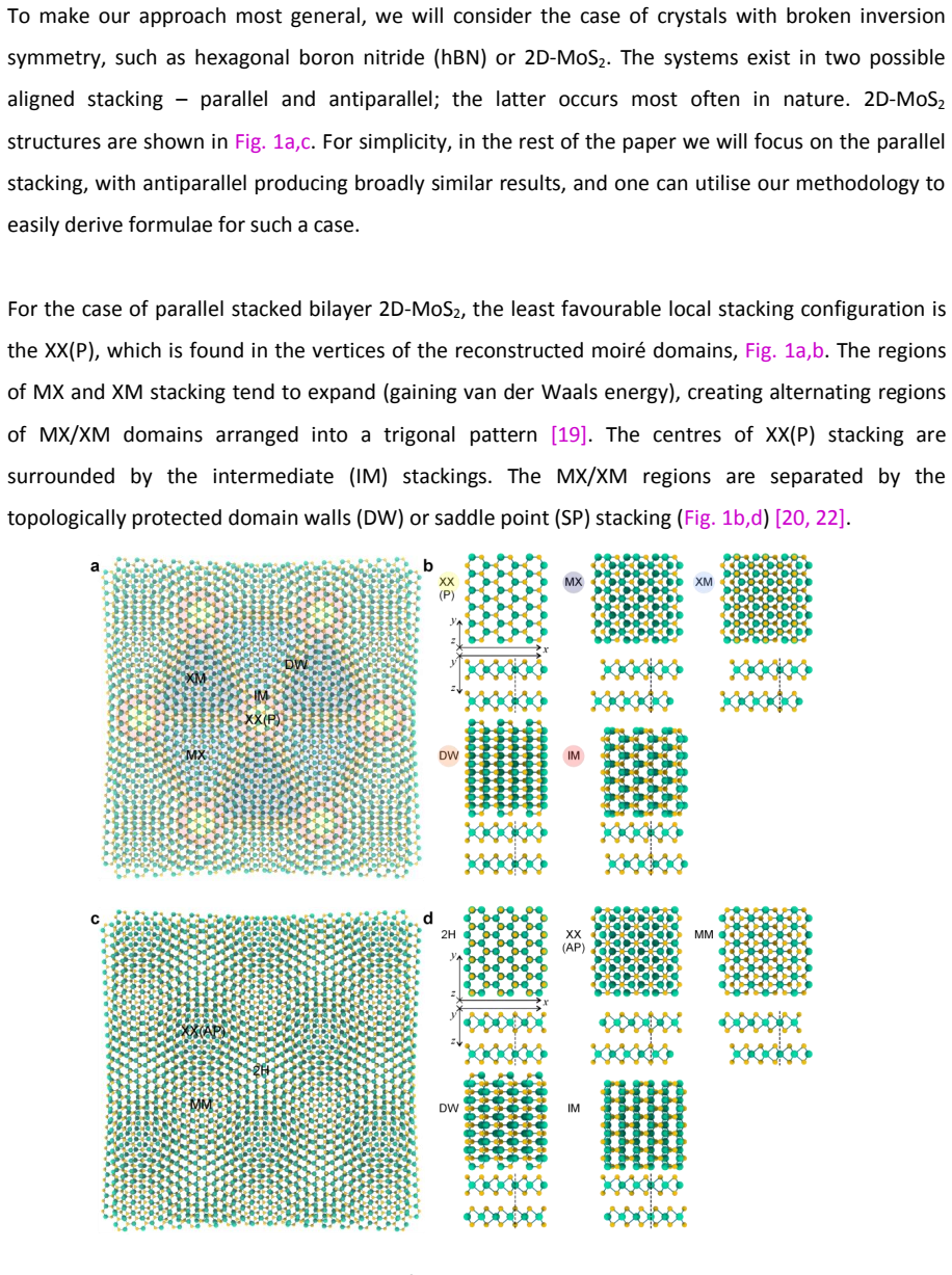
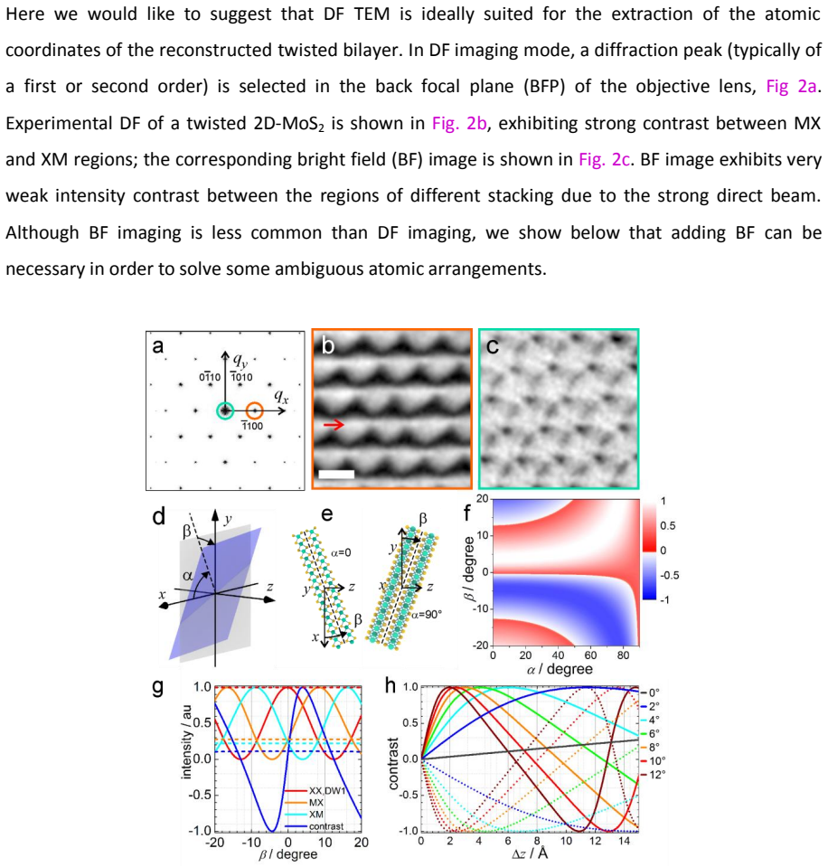
Reconstruction of the atomic crystal structure in twisted 2D materials has been demonstrated to be responsible for multiple exciting phenomena in van der Waals heterostructures, from the appearance of flat bands in twisted bilayer graphene to Wigner crystallization in transition metal dichalcogenides (TMDs). However, there are still no experimental methods for accessing the 3D atomic distributions nor models that describe the exact atomic shifts in such reconstructed structures, which significantly impedes the development of the field. Dark field (DF) transmission electron microscopy (TEM) has been conventionally employed to visualize the local in-plane atomic displacements. Here we expand this method to obtain a full description of the reconstructed atomic systems and demonstrate the quantitative relations between the local stacking and the intensity in the DF image. We show how local 3D atomic displacements and the interlayer distance can be extracted from a DF image.
Editorial analysis
A structured set of objections, weighed in public.
Referee Report
Summary. The manuscript claims that dark field TEM, conventionally used to visualize local in-plane atomic displacements in twisted bilayer 2D crystals, can be expanded to extract full 3D atomic displacements and interlayer distance via demonstrated quantitative relations between local stacking configuration and DF image intensity.
Significance. If the quantitative mapping from stacking to intensity holds after proper controls, the work would address a clear experimental gap by enabling access to 3D atomic distributions in reconstructed van der Waals heterostructures. This could support more accurate modeling of atomic shifts and advance studies of phenomena such as flat bands in twisted bilayer graphene and Wigner crystallization in TMDs. The approach builds directly on established DF-TEM methods without introducing new fitted parameters or ad-hoc entities.
major comments (2)
- [Abstract] Abstract: the central claim that local 3D displacements and interlayer distance 'can be extracted from a DF image' rests on the assumption that DF intensity is quantitatively and uniquely fixed by local stacking. No validation against dynamical scattering, thickness variations, or orientation effects is indicated, yet these are known to produce intensity variations independent of in-plane registry in bilayer DF-TEM.
- [Results/Methods] The manuscript provides no error analysis, simulation benchmarks, or experimental controls to confirm that post-hoc choices do not affect the extracted displacements, leaving the soundness of the quantitative relations unverified.
minor comments (1)
- [Abstract] Abstract: consider adding a sentence specifying the 2D materials or twist angles used in the demonstration to make the scope concrete.
Simulated Author's Rebuttal
We thank the referee for their careful reading and constructive comments, which help strengthen the manuscript. We address each major comment below and indicate planned revisions.
read point-by-point responses
-
Referee: [Abstract] Abstract: the central claim that local 3D displacements and interlayer distance 'can be extracted from a DF image' rests on the assumption that DF intensity is quantitatively and uniquely fixed by local stacking. No validation against dynamical scattering, thickness variations, or orientation effects is indicated, yet these are known to produce intensity variations independent of in-plane registry in bilayer DF-TEM.
Authors: We acknowledge that the abstract claim relies on the intensity being fixed by local stacking under the kinematic approximation used for thin 2D samples. The manuscript derives the relations from this framework but does not explicitly validate against dynamical effects. In revision we will add a dedicated section with multislice simulations benchmarking intensity variations due to dynamical scattering, thickness, and orientation, plus quantitative error bounds on the extracted displacements. revision: yes
-
Referee: [Results/Methods] The manuscript provides no error analysis, simulation benchmarks, or experimental controls to confirm that post-hoc choices do not affect the extracted displacements, leaving the soundness of the quantitative relations unverified.
Authors: We agree the original text lacks explicit error analysis and controls. The revision will incorporate error propagation from measured intensities to 3D positions, simulation benchmarks on model reconstructed structures, and discussion of experimental controls (e.g., thickness series) to demonstrate robustness against processing choices. revision: yes
Circularity Check
No circularity: experimental mapping from DF intensity to 3D structure is presented as empirical demonstration, not self-referential fit or definition
full rationale
The paper claims to demonstrate quantitative relations between local stacking and DF image intensity, then extract 3D displacements and interlayer distance from that mapping. No equations, self-citations, or ansatzes are identified that reduce the extracted quantities to parameters fitted from the same data by construction, nor any uniqueness theorem imported from the authors' prior work. The derivation chain is self-contained as an imaging technique whose validity rests on external validation of the intensity-stacking relation rather than internal redefinition.
Axiom & Free-Parameter Ledger
axioms (1)
- domain assumption DF TEM intensity is quantitatively determined by local stacking configuration
Reference graph
Works this paper leans on
-
[1]
E. J. Kirkland, Advanced Computing in Electron Microscopy (Springer, 2010)
2010
-
[2]
Practical algorithms for simulation and reconstruction of digital in-line holograms,
T. Latychevskaia, and H.-W. Fink, "Practical algorithms for simulation and reconstruction of digital in-line holograms," Appl. Opt. 54, 2424–2434 (2015)
2015
-
[3]
Atomic and electronic reconstruction at the van der Waals interface in twisted bilayer graphene,
H. Yoo et al. , "Atomic and electronic reconstruction at the van der Waals interface in twisted bilayer graphene," Nature Mater. 18, 448–453 (2019)
2019
discussion (0)
Sign in with ORCID, Apple, or X to comment. Anyone can read and Pith papers without signing in.