REVIEW 3 major objections 5 minor 1 cited by
Deep Active Lesion Segmentation
T0 review · 3 major / 5 minor · reviewed 2026-08-14 · deepseek-v4-flash
Pith's one-line read Deep Active Lesion Segmentation couples a CNN encoder-decoder with a level-set active contour model, using learned per-pixel parameter maps to initialize and tune the contour, and reports significantly more accurate lesion boundaries than…
desk verdict Solid CNN+level-set hybrid with a new multi-organ dataset, but the per-organ significance claims exceed what the test sizes can support. read the letter →
The pith
A machine-rendered reading of the paper's core claim, the machinery that carries it, and where it could break.
The reading
What carries the argument
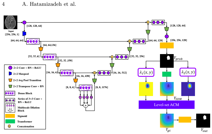
The load-bearing mechanism is a per-pixel-parameterized level-set energy functional, a generalization of the Chan-Vese active contour energy in which the scalar constants $\lambda_1$ and $\lambda_2$ become functions $\lambda_1(x,y)$ and $\lambda_2(x,y)$ over the image domain. An encoder-decoder CNN with dense blocks and a multiscale dilated-convolution block produces the initial probability map $Y_{\mathrm{prob}}$; the Transformer converts it into the signed distance map that initializes the contour, and the exponential formulas above convert it into the parameter maps. During evolution, these local weights let the contour bend precisely to irregular, low-contrast lesion boundaries, which is the mechanism the paper credits for its boundary accuracy.
What would settle it
Run DALS and its CNN backbone on repeated random splits or k-fold cross-validation of each MLS organ subset; if the Dice advantage shrinks or the Wilcoxon p-value rises above 0.05 in any organ, the claimed significant boundary improvement is not supported.
Extended reading notes
Core claim
The central claim is that lesion segmentation can be made both fully automatic and boundary-precise by letting a CNN supply the initial contour and the spatially varying weights of a level-set active contour model. Concretely, the CNN outputs a probability map $Y_{\mathrm{prob}}(x,y)$; a transformer converts it into a signed distance map $\varphi(x,y,0)$ that seeds the contour, and the same map defines per-pixel parameters $\lambda_1(x,y)=\exp\big((2-Y_{\mathrm{prob}})/(1+Y_{\mathrm{prob}})\big)$ and $\lambda_2(x,y)=\exp\big((1+Y_{\mathrm{prob}})/(2-Y_{\mathrm{prob}})\big)$ in the energy functional. The contour then evolves by the Euler-Lagrange PDE of a localized Chan-Vese energy, so each boundary point carries its own interior and exterior weighting. On the paper's Multiorgan Lesion Segmentation dataset, DALS reports higher Dice and boundary metrics than U-Net, a manually initialized level-set model, and its own CNN backbone, and the authors state that the improvements are statistically significant.
Load-bearing premise
The load-bearing premise is that the 10-percent held-out test subsets of the MLS dataset, as few as nine lung images, are representative enough to support the reported significance given that DALS's confidence intervals overlap with its own CNN backbone on several organs.
Editorial extensions
If this is right
- Lesion segmentation becomes fully automatic: no manual contour initialization and no hand-picked active-contour weights, because the network supplies both.
- Boundary quality should improve most where intensity contrast is low or the lesion edge is irregular, exactly the cases pure CNNs handle poorly.
- The same architecture can be retrained for other organs and modalities without changing the level-set machinery.
- The method should remain usable with small training sets, because the CNN only needs to produce a coarse localization and parameter maps rather than a pixel-perfect boundary.
Reading between the lines
- A natural ablation not reported in the paper: replace the learned $\lambda_1(x,y)$ and $\lambda_2(x,y)$ maps with their spatial means while keeping the same initialization; if boundary metrics degrade but Dice does not, the parameter maps are doing the boundary work.
- Because the exponential transforms amplify $Y_{\mathrm{prob}}$ values near 0 and 1, a poorly calibrated probability map could produce extreme local weights; measuring calibration per organ would predict where DALS is most likely to fail.
- The same pipeline should transfer to non-radiology segmentation tasks with irregular boundaries, such as ultrasound lesions or cells, since the level-set evolution is agnostic to image modality.
- The significance claim could be stress-tested by repeated cross-validation; the reported 10 percent test splits leave as few as nine lung images, and several confidence intervals overlap with the backbone CNN.
Signed reviews
Editorial analysis
A structured set of objections, weighed in public.
Referee Report
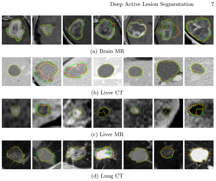
Summary. The paper introduces Deep Active Lesion Segmentation (DALS), a framework combining a fully convolutional encoder-decoder with a level-set active contour model (ACM). The CNN predicts a lesion probability map that initializes the signed distance function and also produces per-pixel parameter maps λ1(x,y) and λ2(x,y) used in a localized Chan-Vese style energy functional. The method is evaluated on a new Multiorgan Lesion Segmentation (MLS) dataset with brain MR, lung CT, liver CT, and liver MR images, comparing against U-Net, a manually initialized level-set ACM, and the authors' CNN backbone. The authors report improved Dice, Hausdorff distance, and Boundary F1 scores, and claim statistically significant superiority via Wilcoxon paired tests.
Significance. If the reported results hold, the work makes a useful contribution to medical image segmentation by demonstrating a practical method for learning per-pixel ACM energy parameters from a CNN, moving beyond scalar or hand-tuned λ values. The release of source code and the introduction of the MLS dataset are positive aspects that support reproducibility and further research. The central architectural idea is plausible and the paper includes a comparison against a strong U-Net baseline, which lends credibility to the approach.
major comments (3)
- [Section 4, Table 2] The statistical support for the headline claim of 'significantly more accurate boundaries' is not established. The paper reports Wilcoxon paired test p-values of p<0.001 against U-Net and the level-set baseline, and p<0.005 against the CNN backbone, but with the stated split (85/10/5) the lung CT test set consists of roughly 9 images and the liver CT test set of roughly 11 images. For a paired Wilcoxon test with n=9, the smallest attainable two-sided p-value is 2/2^9 = 0.0039, so p<0.001 cannot be correct if the test was performed per organ. If the test was pooled across organs, then it does not independently support the per-dataset claim 'in all datasets' made in the same paragraph. In addition, the confidence intervals reported in Table 2 for DALS and the CNN backbone overlap on lung CT (Dice 0.869 ± 0.092 vs 0.822 ± 0.094) and on liver CT (0.846 ± 0.081 vs 0.801 ± 0.159), which further undermines the per-organ significance claim. The authors must specify the exact test procedure, provide per-organ p-values or confidence intervals of the differences, and correct the reported p-values if they are unattainable.
- [Section 3, data split] The evaluation rests on a single 10% test split per organ, with no repeated resampling, cross-validation, or description of patient-level splitting. For the lung CT subset, the test set is only about 8–9 images, which is too small to support general claims about 'all datasets' without additional evidence. The paper should report the exact number of test images per organ, clarify whether images from the same patient appear across train/test splits, and ideally provide leave-one-out or repeated random sub-sampling results to demonstrate that the reported gains are not an artifact of a particular split.
- [Section 4, Algorithm Comparison] The manually initialized level-set ACM baseline is unsupervised: it does not use the training labels, while DALS is fully supervised. Comparing a supervised method against an unsupervised baseline does not isolate the contribution of the per-pixel parameter functions, because the baseline does not receive any task-specific tuning. A fairer ablation would either train the same backbone with scalar λ constants (for example, by integrating the level-set evolution into the training loop, as in the cited deep level-set works) or at least tune a scalar λ on the validation set and then compare. Without such a control, the claim that the learned parameter maps are responsible for the improvement is not convincingly supported.
minor comments (5)
- [Abstract] The sentence 'we introduce Deep Active Lesion Segmentation (DALS), a fully automated segmentation framework that leverages...' contains a grammatical issue ('that leverages' is split from its antecedent by 'framework'); consider revising for readability.
- [Section 2.3, Eq. (3)] The exponential forms for λ1 and λ2 are presented without motivation for the specific formulas. A brief explanation of why the chosen exponential functions are appropriate, or a reference to the prior work that used them, would help readers understand the design choice.
- [Table 2] In the Liver CT row, the Hausdorff CI for the CNN Backbone is listed as 1.6, which is an order of magnitude larger than all other CI values in the table and appears to be a typographical error. Please check whether this should be 0.16 or another value.
- [Section 4, statistics] The Wilcoxon test description does not state whether the test is one-sided or two-sided, how ties are handled, or whether any multiple-comparison correction was applied across the four organs and three comparisons. Please provide these details.
- [Figure 2] The tensor dimensions shown in Figure 2 are not fully consistent: for example, the input is [256,256,1] but the first feature block is [128,128,64], and the concatenated feature maps before the multiscale dilation block have different listed sizes. Clarify the notation to show how these dimensions are obtained.
Circularity Check
No significant circularity: the method is evaluated against held-out ground truth and an external U-Net baseline, and the CNN-to-ACM coupling is an architectural choice rather than a fitted-input-as-prediction design.
full rationale
The paper's central claim is that DALS, which combines a CNN backbone with a level-set ACM, yields more accurate lesion boundaries than U-Net, a manually initialized level-set ACM, and its own CNN backbone. The evaluation is performed on held-out test images against expert ground truth, and the comparisons include an external baseline (U-Net). The lambda parameter maps in Eq. (3) are computed from the CNN probability map Yprob, which was trained with a Dice loss against ground truth; this means the ACM refinement is coupled to the CNN prediction rather than being fully independent. However, this is a design coupling, not a definitional circularity: the paper does not define the segmentation accuracy in terms of the fitted parameters, nor does it rename a fitted quantity as a prediction. The extension of Hoogi et al. [3] is cited for the form of the lambda functions, but the formula is stated explicitly in Eq. (3), so the argument does not reduce to an unverified self-citation. The reported Wilcoxon p-values may be statistically questionable given the small per-organ test sizes, but that is a correctness or statistical-power concern, not evidence of circularity. No load-bearing step in the derivation chain reduces by construction to its own inputs, and no fitted parameter is relabeled as a prediction. Therefore, the paper receives a circularity score of 0.
Assumptions & free parameters
free parameters (2)
- mu (length penalty) =
0.1
- lambda map exponential formula =
exponents (2-Y)/(1+Y) and (1+Y)/(2-Y)
assumptions (5)
- standard math Chan-Vese uniform internal energy model
- standard math Lankton localized region integration
- domain assumption Numerical time integration of the level-set PDE converges to a useful minimum
- domain assumption Manual ground truth annotations are accurate and consistent
- ad hoc to paper The CNN probability map Yprob is sufficiently accurate to initialize and weight the ACM
Cite this review
Pith. "Pith review of Deep Active Lesion Segmentation." pith.science (2026). https://pith.science/paper/LCHGXQTG
@misc{pith2026190806933,
  author       = {Pith},
  title        = {Pith review of: Deep Active Lesion Segmentation},
  year         = {2026},
  howpublished = {\url{https://pith.science/paper/LCHGXQTG}},
  note         = {Machine review of arXiv:1908.06933}
}
abstract
Lesion segmentation is an important problem in computer-assisted diagnosis that remains challenging due to the prevalence of low contrast, irregular boundaries that are unamenable to shape priors. We introduce Deep Active Lesion Segmentation (DALS), a fully automated segmentation framework for that leverages the powerful nonlinear feature extraction abilities of fully Convolutional Neural Networks (CNNs) and the precise boundary delineation abilities of Active Contour Models (ACMs). Our DALS framework benefits from an improved level-set ACM formulation with a per-pixel-parameterized energy functional and a novel multiscale encoder-decoder CNN that learns an initialization probability map along with parameter maps for the ACM. We evaluate our lesion segmentation model on a new Multiorgan Lesion Segmentation (MLS) dataset that contains images of various organs, including brain, liver, and lung, across different imaging modalities---MR and CT. Our results demonstrate favorable performance compared to competing methods, especially for small training datasets. Source code : $\text{https://github.com/ahatamiz/dals}$
Figures


Figures from the paper (2 more)

Forward citations
Cited by 1 Pith paper
-
End-to-End Boundary Aware Networks for Medical Image Segmentation
A boundary-aware CNN with a dedicated edge stream and edge losses improves tumor-core segmentation on 2D slices of BraTS 2018 compared to U-Net and V-Net, though the evaluation has internal inconsistencies.
Reference graph
Works this paper leans on
-
[1]
IEEE Transactions on Image Processing 10(2), 266--277 (2001)
Chan, T.F., Vese, L.A.: Active contours without edges. IEEE Transactions on Image Processing 10(2), 266--277 (2001)
work page 2001
-
[2]
arXiv preprint arXiv:1905.12120 (2019)
Hatamizadeh, A., Hosseini, H., Liu, Z., Schwartz, S.D., Terzopoulos, D.: Deep dilated convolutional nets for the automatic segmentation of retinal vessels. arXiv preprint arXiv:1905.12120 (2019)
arXiv 2019
-
[3]
IEEE Transactions on Medical Imaging 36(3), 781--791 (2017)
Hoogi, A., Subramaniam, A., Veerapaneni, R., Rubin, D.L.: Adaptive estimation of active contour parameters using convolutional neural networks and texture analysis. IEEE Transactions on Medical Imaging 36(3), 781--791 (2017)
work page 2017
- [4]
-
[5]
In: Deep Learning in Medical Image Analysis, Lecture Notes in Computer Science, vol
Imran, A.A.Z., Hatamizadeh, A., Ananth, S.P., Ding, X., Terzopoulos, D., Tajbakhsh, N.: Automatic segmentation of pulmonary lobes using a progressive dense V -network. In: Deep Learning in Medical Image Analysis, Lecture Notes in Computer Science, vol. 11045, pp. 282--290. Springer (2018)
work page 2018
-
[6]
International Journal of Computer Vision 1(4), 321--331 (1988)
Kass, M., Witkin, A., Terzopoulos, D.: Snakes: Active contour models. International Journal of Computer Vision 1(4), 321--331 (1988)
work page 1988
-
[7]
IEEE Transactions on Image Processing 17(11), 2029--2039 (2008)
Lankton, S., Tannenbaum, A.: Localizing region-based active contours. IEEE Transactions on Image Processing 17(11), 2029--2039 (2008)
work page 2008
- [8]
Show all 11 references
-
[9]
In: LNCS vol
Ronneberger, O., Fischer, P., Brox, T.: U-net: Convolutional networks for biomedical image segmentation. In: LNCS vol. 9351 (Proc. MICCAI). pp. 234--241 (2015)
2015
-
[10]
, " * write output.state after.block = add.period write newline
ENTRY address author booktitle chapter edition editor eid howpublished institution journal key month note number organization pages publisher school series title type url volume year label extra.label sort.label short.list INTEGERS output.state before.all mid.sentence after.se...
-
[11]
write newline
" write newline "" before.all 'output.state := FUNCTION n.dashify 't := "" t empty not t #1 #1 substring "-" = t #1 #2 substring "--" = not "--" * t #2 global.max substring 't := t #1 #1 substring "-" = "-" * t #2 global.max substring 't := while if t #1 #1 substring * t #2 gl...
Reviewed August 14, 2026 · model on record in the stance chip above.
Discussion (0). Continue with ORCID to comment.