REVIEW 3 major objections 4 minor 97 references
Effectiveness of self-supervised learning in medical imaging is decided by the fit between pretext objective, imaging modality, and clinical task—not by any single method.
Reviewed by Pith at T0; open to challenge. T0 means a machine referee read the full paper against a public rubric. the ladder, T0–T4 →
A task-oriented review of medical-image SSL arguing that pretext tasks should be chosen to match downstream tasks and modalities, not used one-size-fits-all.
T0 review reviewed 2026-08-02 challenge →
load-bearing objection Useful task-alignment review with a plausible central claim, but the evidence table is qualitative and the manuscript has auditability issues that should be fixed before publication. the 3 major comments →
Task-Aligned Self-Supervised Learning for Medical Image Analysis: A Task-Oriented Review with Practical Design Guidelines
The pith
A machine-rendered reading of the paper's core claim, the machinery that carries it, and where it could break.
The reading
Core claim
The paper's central discovery—offered as a synthesis of the surveyed literature—is that SSL effectiveness in medical imaging is context-dependent, governed by the match among pretext objective, imaging modality, and downstream task. It encodes this as a task-alignment matrix: instance- and patient-level contrastive learning aligns strongly with classification but weakly with reconstruction and regression; local/voxel-level contrastive, non-contrastive and predictive, and generative/reconstruction objectives align with segmentation and detection; hybrid methods balance global and local information at the cost of training complexity. The paper also identifies modality-specific patterns (volume
What carries the argument
The load-bearing device is the task-alignment matrix (Table 3), a qualitative map of alignment strength between four SSL pretext-task families and five downstream clinical objectives. The review also provides a literature-derived taxonomy dividing pretext tasks into contrastive (instance-level, patient-level, local/voxel, cross-modal, acquisition-based), non-contrastive and predictive (self-distillation, redundancy reduction, spatial/anatomical prediction, temporal prediction), generative and reconstruction-based (masked modeling, context restoration, cross-modal synthesis, colorization), and hybrid learning. The matrix does the work of the argument: it turns the collected studies into a str
Load-bearing premise
The central claim rests on the assumption that the 75 selected studies fairly represent the field and that the authors' qualitative alignment ratings (Table 3) accurately summarize the true trends, since no effect-size meta-analysis or fully specified search protocol backs the ratings.
What would settle it
A systematic meta-analysis of the 75 reviewed studies, extracting effect sizes (e.g., Dice or AUC improvements over supervised baselines) grouped by pretext family, downstream task, and modality, would settle the claim: if the alignment interaction pattern in Table 3 does not appear in the aggregated numbers, the central conclusion collapses. A cheaper check: a controlled benchmark with fixed architecture, data, and compute that varies pretext tasks across classification, segmentation, and detection tasks and shows the claimed 'strong/moderate/weak' ordering does not reproduce.
If this is right
- SSL method selection in medical imaging should be driven by a three-way fit—pretext objective, imaging modality, and clinical target—rather than by benchmark popularity or direct transfer from natural-image SSL.
- Misaligned objectives risk negative transfer: rotation-invariant contrastive learning can erase orientation cues that are diagnostic in cardiac MRI, and high masking ratios can remove small lesions in CT and dental radiographs.
- In low-label and few-shot regimes, well-aligned SSL pretraining can match or exceed fully supervised training with full labels—for example, 86% accuracy in liver-view classification with one labeled image per class.
- Hybrid objectives are the most general-purpose choice but carry higher training complexity and computational cost, making them less practical where resources are constrained.
- Evaluation of SSL in medical imaging should move beyond average downstream accuracy to measure preservation of diagnostically relevant local structures, robustness, and out-of-distribution generalization.
Where Pith is reading between the lines
- A natural extension the review leaves implicit: the alignment matrix could be operationalized as a decision rule—task type leading to a preferred pretext family, then to modality-specific masking and augmentation budgets—giving practitioners a concrete starting point without re-running the survey.
- The qualitative alignment ratings could be tested directly by a controlled benchmark that holds architecture, data, and compute fixed and varies pretext family across classification, segmentation, and detection; if the interaction pattern in Table 3 fails to reproduce, the review's central synthesis would be weakened.
- The review's world-model suggestion—predicting future anatomical states rather than masked pixels—points to a testable extension: longitudinal medical data could train prediction-based pretext tasks that should outperform static reconstruction tasks on progression-tracking endpoints, a claim the paper mentions but does not evaluate.
- Extracting reported effect sizes from the 75 studies and grouping them by pretext family, task, and modality would allow a quantitative check of the 'strong/moderate/weak' alignment ratings; the review itself does not perform such a meta-analysis.
Editorial analysis
A structured set of objections, weighed in public.
Referee Report
Summary. This manuscript is a task-oriented review of self-supervised learning (SSL) for medical image analysis. The authors analyze 75 studies (abstract states 78) published 2017–2025, organizing them into four pretext-task families: contrastive, non-contrastive/predictive, generative/reconstruction-based, and hybrid. The central claim is that SSL effectiveness is governed by the match among pretext objective, imaging modality, and downstream task, rather than by any single strategy. The paper provides a taxonomy, a qualitative task-alignment matrix (Table 3), analyses of modality and label-regime effects, and practical design guidelines (Table 4). The review explicitly declines a quantitative meta-analysis because of heterogeneity.
Significance. If the task-alignment thesis is correct, this review would provide valuable practical guidance for selecting SSL methods in medical imaging, moving beyond generic 'SSL works' conclusions. The paper's strengths include a broad coverage of the literature, a clear four-family taxonomy, a design-guideline table that is directly actionable, and an explicit acknowledgment of common pitfalls such as masking-ratio sensitivity and modality mismatch. However, the central claim rests on qualitative synthesis rather than measurable effect sizes, and the current manuscript has an incomplete search protocol and internal inconsistencies. As a review contribution, the work is potentially useful, but its evidence base needs to be made auditable and its claims more carefully calibrated to the strength of the underlying studies.
major comments (3)
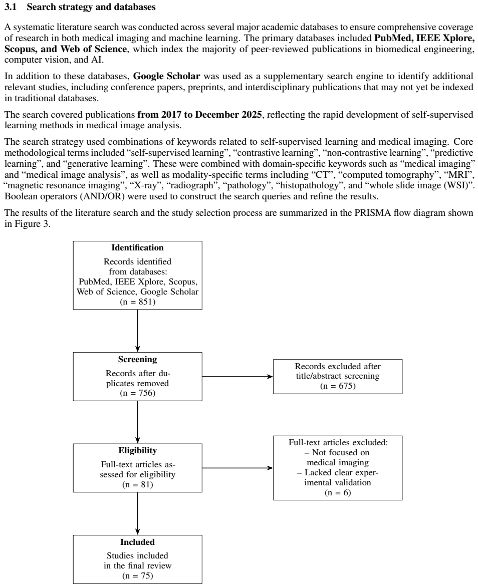
- [Section 2, literature selection] The search protocol is incomplete: the manuscript contains the literal placeholder '[insert databases used, e.g., PubMed, IEEE Xplore, ScienceDirect, SpringerLink, Scopus, Web of Science, and arXiv]'. Without an explicit, completed search strategy, the representativeness of the 75-study corpus cannot be assessed, and the review's central qualitative claim is not auditable. In addition, the abstract reports '78 studies' while Section 2 and Table 2 report 75. These inconsistencies must be resolved and the protocol described in full.
- [Table 3, Section 5.2] The task-alignment matrix is the principal evidence for the paper's central claim, yet the ratings are qualitative author judgments with no reported effect sizes, no per-cell number of supporting studies, and no explicit inclusion criteria for rating assignment. Because the underlying studies are between-study comparisons with heterogeneous datasets, backbones, and evaluation protocols, the observed pattern (contrastive strong for classification, spatial/generative strong for segmentation) could reflect benchmark selection or architectural confounds rather than true task-alignment. Section 7 correctly notes that 'it is difficult to isolate the true contributions of SSL from other factors,' but this caveat is not applied to Table 3. At minimum, each rating should be justified with representative citations and a statement of how disagreements or heterogeneous results were resolved.
- [Section 5.5.2 (negative transfer)] The claim that misaligned objectives can cause negative transfer is presented as an established finding, but the cited evidence is anecdotal. For example, the ~5–6% performance drop in [37] refers to concatenating modalities without modeling relationships, not necessarily to a misaligned pretext objective; the masking-ratio sensitivity in [24,66] is a hyperparameter effect within a single method. The abstract's stronger statement that misaligned objectives 'can cause negative transfer through shortcut learning on acquisition signatures or augmentation that erases diagnostic signal' goes beyond what these examples support. I recommend framing negative transfer as a plausible hypothesis with preliminary support, and specifying which studies directly demonstrate it under controlled comparisons.
minor comments (4)
- [Section 4, introductory sentence] Typo: 'Sectio' should be 'Section'.
- [Section 5.2.3] Citation inconsistency: the text says 'Almalki and Latecki [69] successfully aligned SimMIM with both teeth numbering and restoration detection,' but reference [69] is the dental-MAE mesh modeling paper; reference [66] is the SimMIM dental radiograph paper. Please correct the citation.
- [Section 9, Conclusion] Typo: 'V olumetric' should be 'Volumetric'.
- [Section 1, introduction, second paragraph] There is a duplicate sentence: 'SSL has become an important strategy...' is immediately followed by a nearly identical sentence with slightly different wording. Please consolidate.
Circularity Check
No significant circularity: the task-alignment conclusion is a qualitative synthesis of 75 external studies, not a derivation from fitted parameters, self-citations, or definitions.
full rationale
This is a narrative review, not a derivation. The central claim—that SSL effectiveness is governed by the match among pretext objective, modality, and downstream task—is supported by reference to 75 external studies, and Table 3 is explicitly labeled as a qualitative synthesis of recurring trends, not as a fitted or independently predicted quantity. There are no equations, no parameter fits, and no author self-citations in the reference list; the load-bearing evidence is external. The review's own Section 7 acknowledges that 'it is difficult to isolate the true contributions of SSL from other factors' and that standardized protocols are needed, which is a correctness/evidence limitation rather than a circularity. Section 2 still contains a placeholder ('[insert databases used, e.g., PubMed...]') and the abstract/body study counts differ (78 vs 75), undermining auditability, but neither makes the argument circular. The only self-referential aspect is that the taxonomy and Table 3 ratings are constructed from the same studies they summarize; however, the paper presents this explicitly as a qualitative review synthesis, not as a prediction derived from a fitted input, so it does not reduce to its own inputs by construction.
Axiom & Free-Parameter Ledger
axioms (3)
- domain assumption The 75 reviewed primary studies accurately report their experimental results and comparisons.
- ad hoc to paper The four-family taxonomy (contrastive, non-contrastive/predictive, generative, hybrid) is an appropriate and non-overlapping partition of pretext tasks.
- domain assumption Table 3's qualitative alignment ratings fairly summarize the trend across heterogeneous studies.
Cite this review
Pith. "Pith review of Task-Aligned Self-Supervised Learning for Medical Image Analysis: A Task-Oriented Review with Practical Design Guidelines." pith.science (2026). https://pith.science/paper/LKNWGSQK
@misc{pith2026260523995,
  author       = {Pith},
  title        = {Pith review of: Task-Aligned Self-Supervised Learning for Medical Image Analysis: A Task-Oriented Review with Practical Design Guidelines},
  year         = {2026},
  howpublished = {\url{https://pith.science/paper/LKNWGSQK}},
  note         = {Machine review of arXiv:2605.23995}
}
abstract
Self-supervised learning (SSL) is increasingly used in medical image analysis to reduce dependence on costly expert annotations by learning transferable representations from unlabeled data. However, SSL performance depends not only on model architecture but also on whether the self-supervised objective preserves the information required by the downstream clinical task. This review presents a task-oriented synthesis of SSL methods for medical imaging, focusing on how the design of the self-supervised objective interacts with imaging modality, label availability, and downstream performance. We analyze $78$ studies published from 2017 to 2025 and organize them into four paradigms: contrastive, non-contrastive and predictive, generative and reconstruction-based, and hybrid learning. Rather than cataloging methods chronologically, we examine how these paradigms support classification, segmentation, detection, reconstruction, and regression. The evidence suggests that effectiveness is governed by the match among objective, modality, and downstream task rather than by any single strategy. Contrastive objectives favor global discriminative representations suited to classification but may underrepresent localized pathology, whereas spatial-prediction, masked-modeling, and reconstruction objectives better preserve anatomical structure for segmentation and dense prediction. Critically, misaligned objectives can cause negative transfer through shortcut learning on acquisition signatures or augmentation that erases diagnostic signal rather than merely weaker gains. SSL is most beneficial in low-label regimes, but its effectiveness depends on modality-aware augmentation, pathology-preserving corruption, and clinically meaningful evaluation. We conclude with practical design guidelines and open challenges for clinically aligned SSL.
Figures


Reference graph
Works this paper leans on
-
[1]
Preparing medical imaging data for machine learning.Radiology, 295(1):4–15, 2020
Martin J Willemink, Wojciech A Koszek, Cailin Hardell, Jie Wu, Dominik Fleischmann, Hugh Harvey, Les R Folio, Ronald M Summers, Daniel L Rubin, and Matthew P Lungren. Preparing medical imaging data for machine learning.Radiology, 295(1):4–15, 2020
2020
-
[2]
Embracing imperfect datasets: A review of deep learning solutions for medical image segmentation.Medical image analysis, 63:101693, 2020
Nima Tajbakhsh, Laura Jeyaseelan, Qian Li, Jeffrey N Chiang, Zhihao Wu, and Xiaowei Ding. Embracing imperfect datasets: A review of deep learning solutions for medical image segmentation.Medical image analysis, 63:101693, 2020
2020
-
[3]
Self-supervised learning methods and applications in medical imaging analysis: A survey.PeerJ Computer Science, 8:e1045, 2022
Saeed Shurrab and Rehab Duwairi. Self-supervised learning methods and applications in medical imaging analysis: A survey.PeerJ Computer Science, 8:e1045, 2022
2022
-
[4]
Self-supervised visual feature learning with deep neural networks: A survey
Longlong Jing and Yingli Tian. Self-supervised visual feature learning with deep neural networks: A survey. IEEE transactions on pattern analysis and machine intelligence, 43(11):4037–4058, 2020
2020
-
[5]
S Kevin Zhou, Hayit Greenspan, Christos Davatzikos, James S Duncan, Bram Van Ginneken, Anant Madabhushi, Jerry L Prince, Daniel Rueckert, and Ronald M Summers. A review of deep learning in medical imaging: Imaging traits, technology trends, case studies with progress highlights, and future promises.Proceedings of the IEEE, 109(5):820–838, 2021. 24
2021
-
[6]
Masked autoencoders are scalable vision learners
Kaiming He, Xinlei Chen, Saining Xie, Yanghao Li, Piotr Dollár, and Ross Girshick. Masked autoencoders are scalable vision learners. InProceedings of the IEEE/CVF conference on computer vision and pattern recognition, pages 16000–16009, 2022
2022
-
[7]
A simple framework for contrastive learning of visual representations
Ting Chen, Simon Kornblith, Mohammad Norouzi, and Geoffrey Hinton. A simple framework for contrastive learning of visual representations. InInternational conference on machine learning, pages 1597–1607. PmLR, 2020
2020
-
[8]
Spyros Gidaris, Praveer Singh, and Nikos Komodakis. Unsupervised representation learning by predicting image rotations.arXiv preprint arXiv:1803.07728, 2018
Pith/arXiv arXiv 2018
-
[9]
Unsupervised learning of visual representations by solving jigsaw puzzles
Mehdi Noroozi and Paolo Favaro. Unsupervised learning of visual representations by solving jigsaw puzzles. In European conference on computer vision, pages 69–84. Springer, 2016
2016
-
[10]
Demystifying contrastive self-supervised learning: Invariances, augmentations and dataset biases.Advances in Neural Information Processing Systems, 33:3407–3418, 2020
Senthil Purushwalkam and Abhinav Gupta. Demystifying contrastive self-supervised learning: Invariances, augmentations and dataset biases.Advances in Neural Information Processing Systems, 33:3407–3418, 2020
2020
-
[11]
Multimodal mr synthesis via modality-invariant latent representation.IEEE transactions on medical imaging, 37(3):803–814, 2017
Agisilaos Chartsias, Thomas Joyce, Mario Valerio Giuffrida, and Sotirios A Tsaftaris. Multimodal mr synthesis via modality-invariant latent representation.IEEE transactions on medical imaging, 37(3):803–814, 2017
2017
-
[12]
Medgan: Medical image translation using gans.Computerized medical imaging and graphics, 79:101684, 2020
Karim Armanious, Chenming Jiang, Marc Fischer, Thomas Küstner, Tobias Hepp, Konstantin Nikolaou, Sergios Gatidis, and Bin Yang. Medgan: Medical image translation using gans.Computerized medical imaging and graphics, 79:101684, 2020
2020
-
[13]
Self-supervised learning for medical image classification: a systematic review and implementation guidelines
Shih-Cheng Huang, Anuj Pareek, Malte Jensen, Matthew P Lungren, Serena Yeung, and Akshay S Chaudhari. Self-supervised learning for medical image classification: a systematic review and implementation guidelines. NPJ Digital Medicine, 6(1):74, 2023
2023
-
[14]
A survey of the impact of self-supervised pretraining for diagnostic tasks in medical x-ray, ct, mri, and ultrasound.BMC medical imaging, 24(1):79, 2024
Blake VanBerlo, Jesse Hoey, and Alexander Wong. A survey of the impact of self-supervised pretraining for diagnostic tasks in medical x-ray, ct, mri, and ultrasound.BMC medical imaging, 24(1):79, 2024
2024
-
[15]
Self-supervised visual representation learning for medical image analysis: A comprehensive survey.Transactions on Machine Learning Research, 2024
Siladittya Manna, Saumik Bhattacharya, and Umapada Pal. Self-supervised visual representation learning for medical image analysis: A comprehensive survey.Transactions on Machine Learning Research, 2024
2024
-
[16]
Self-supervised learning in medicine and healthcare.Nature Biomedical Engineering, 6(12):1346–1352, 2022
Rayan Krishnan, Pranav Rajpurkar, and Eric J Topol. Self-supervised learning in medicine and healthcare.Nature Biomedical Engineering, 6(12):1346–1352, 2022
2022
-
[17]
3d self-supervised methods for medical imaging.Advances in neural information processing systems, 33:18158–18172, 2020
Aiham Taleb, Winfried Loetzsch, Noel Danz, Julius Severin, Thomas Gaertner, Benjamin Bergner, and Christoph Lippert. 3d self-supervised methods for medical imaging.Advances in neural information processing systems, 33:18158–18172, 2020
2020
-
[18]
Rubik’s cube+: A self-supervised feature learning framework for 3d medical image analysis.Medical image analysis, 64:101746, 2020
Jiuwen Zhu, Yuexiang Li, Yifan Hu, Kai Ma, S Kevin Zhou, and Yefeng Zheng. Rubik’s cube+: A self-supervised feature learning framework for 3d medical image analysis.Medical image analysis, 64:101746, 2020
2020
-
[19]
Extracting and composing robust features with denoising autoencoders
Pascal Vincent, Hugo Larochelle, Yoshua Bengio, and Pierre-Antoine Manzagol. Extracting and composing robust features with denoising autoencoders. InProceedings of the 25th international conference on Machine learning, pages 1096–1103, 2008
2008
-
[20]
Momentum contrast for unsupervised visual representation learning
Kaiming He, Haoqi Fan, Yuxin Wu, Saining Xie, and Ross Girshick. Momentum contrast for unsupervised visual representation learning. InProceedings of the IEEE/CVF conference on computer vision and pattern recognition, pages 9729–9738, 2020
2020
-
[21]
Dense contrastive learning for self- supervised visual pre-training
Xinlong Wang, Rufeng Zhang, Chunhua Shen, Tao Kong, and Lei Li. Dense contrastive learning for self- supervised visual pre-training. InProceedings of the IEEE/CVF conference on computer vision and pattern recognition, pages 3024–3033, 2021
2021
-
[22]
A survey on self-supervised learning: Recent advances and open problems.Neurocomputing, page 131409, 2025
Jianping Zhang, Lei Yang, Seyed Mahmoud Sajjadi Mohammadabadi, and Feng Yan. A survey on self-supervised learning: Recent advances and open problems.Neurocomputing, page 131409, 2025
2025
-
[23]
Nikolaos Giakoumoglou, Tania Stathaki, and Athanasios Gkelias. A review on discriminative self-supervised learning methods in computer vision.arXiv preprint arXiv:2405.04969, 2024
Pith/arXiv arXiv 2024
-
[24]
Maemc-net: a hybrid self-supervised learning method for predicting the malignancy of solitary pulmonary nodules from ct images.Frontiers in medicine, 12:1507258, 2025
Tianhu Zhao, Yong Yue, Hang Sun, Jingxu Li, Yanhua Wen, Yudong Yao, Wei Qian, Yubao Guan, and Shouliang Qi. Maemc-net: a hybrid self-supervised learning method for predicting the malignancy of solitary pulmonary nodules from ct images.Frontiers in medicine, 12:1507258, 2025
2025
-
[25]
Self supervised contrastive learning for digital histopathology
Ozan Ciga, Tony Xu, and Anne Louise Martel. Self supervised contrastive learning for digital histopathology. Machine learning with applications, 7:100198, 2022
2022
-
[26]
Self-supervised learning for accurate liver view classification in ultrasound images with minimal labeled data
Abder-Rahman Ali, Anthony E Samir, and Peng Guo. Self-supervised learning for accurate liver view classification in ultrasound images with minimal labeled data. Inproceedings of the IEEE/CVF Conference on Computer Vision and Pattern Recognition, pages 3087–3093, 2023. 25
2023
-
[27]
Self-supervised learning leads to improved performance in biparametric prostate mri classification.Computers in Biology and Medicine, 198:111262, 2025
José Guilherme de Almeida, Ana Sofia Castro Verde, Ana Mascarenhas Gaivão, Carlos Bilreiro, Inês Santiago, Joana Ip, Sara Belião, Celso Matos, Manolis Tsiknakis, Kostas Marias, et al. Self-supervised learning leads to improved performance in biparametric prostate mri classification.Computers in Biology and Medicine, 198:111262, 2025
2025
-
[28]
Lingmiao Meng, Liwei Zhao, Xin Yi, and Qingming Yu. Self-supervised contrastive learning for automated segmentation of brain tumor mri images in schizophrenia.International Journal of Computational Intelligence Systems, 17(1):196, 2024
2024
-
[29]
Anuroop Sriram, Matthew Muckley, Koustuv Sinha, Farah Shamout, Joelle Pineau, Krzysztof J Geras, Lea Azour, Yindalon Aphinyanaphongs, Nafissa Yakubova, and William Moore. Covid-19 prognosis via self-supervised representation learning and multi-image prediction.arXiv preprint arXiv:2101.04909, 2021
Pith/arXiv arXiv 2021
-
[30]
Momentum contrastive learning for few-shot covid-19 diagnosis from chest ct images.Pattern recognition, 113:107826, 2021
Xiaocong Chen, Lina Yao, Tao Zhou, Jinming Dong, and Yu Zhang. Momentum contrastive learning for few-shot covid-19 diagnosis from chest ct images.Pattern recognition, 113:107826, 2021
2021
-
[31]
Chess: Chest x-ray pre-trained model via self-supervised contrastive learning.Journal of Digital Imaging, 36(3):902–910, 2023
Kyungjin Cho, Ki Duk Kim, Yujin Nam, Jiheon Jeong, Jeeyoung Kim, Changyong Choi, Soyoung Lee, Jun Soo Lee, Seoyeon Woo, Gil-Sun Hong, et al. Chess: Chest x-ray pre-trained model via self-supervised contrastive learning.Journal of Digital Imaging, 36(3):902–910, 2023
2023
-
[32]
Contrastive self-supervised learning for neurodegenerative disorder classification.Frontiers in Neuroinformatics, 19:1527582, 2025
Vadym Gryshchuk, Devesh Singh, Stefan Teipel, Martin Dyrba, and FTLDNI study groups ADNI, AIBL. Contrastive self-supervised learning for neurodegenerative disorder classification.Frontiers in Neuroinformatics, 19:1527582, 2025
2025
-
[33]
Parcl: Pathology-aware representation contrastive learning for glaucoma classification on fundus images
Junyan Yi, Ying Zheng, Dayong Ding, Jianchun Zhao, and Gang Yang. Parcl: Pathology-aware representation contrastive learning for glaucoma classification on fundus images. In2024 IEEE International Conference on Bioinformatics and Biomedicine (BIBM), pages 3912–3917. IEEE, 2024
2024
-
[34]
Big self-supervised models advance medical image classification
Shekoofeh Azizi, Basil Mustafa, Fiona Ryan, Zachary Beaver, Jan Freyberg, Jonathan Deaton, Aaron Loh, Alan Karthikesalingam, Simon Kornblith, Ting Chen, et al. Big self-supervised models advance medical image classification. InProceedings of the IEEE/CVF international conference on computer vision, pages 3478–3488, 2021
2021
-
[35]
Self-fi: self-supervised learning for disease diagnosis in fundus images.Bioengineering, 10(9):1089, 2023
Toan Duc Nguyen, Duc-Tai Le, Junghyun Bum, Seongho Kim, Su Jeong Song, and Hyunseung Choo. Self-fi: self-supervised learning for disease diagnosis in fundus images.Bioengineering, 10(9):1089, 2023
2023
-
[36]
Self-supervised learning for spinal mris
Amir Jamaludin, Timor Kadir, and Andrew Zisserman. Self-supervised learning for spinal mris. InInternational Workshop on Deep Learning in Medical Image Analysis, pages 294–302. Springer, 2017
2017
-
[37]
Self-supervised feature learning via exploiting multi-modal data for retinal disease diagnosis.IEEE Transactions on Medical Imaging, 39(12):4023– 4033, 2020
Xiaomeng Li, Mengyu Jia, Md Tauhidul Islam, Lequan Yu, and Lei Xing. Self-supervised feature learning via exploiting multi-modal data for retinal disease diagnosis.IEEE Transactions on Medical Imaging, 39(12):4023– 4033, 2020
2020
-
[38]
vox2vec: A framework for self-supervised contrastive learning of voxel-level representations in medical images
Mikhail Goncharov, Vera Soboleva, Anvar Kurmukov, Maxim Pisov, and Mikhail Belyaev. vox2vec: A framework for self-supervised contrastive learning of voxel-level representations in medical images. InInternational Conference on Medical Image Computing and Computer-Assisted Intervention, pages 605–614. Springer, 2023
2023
-
[39]
Multi-encoding contrastive learning for dual-stream self-supervised 3d dental segmentation network.Technologies, 13(9):419, 2025
Tian Ma, Xiaoyuan Wei, Jiechen Zhai, Ziang Zhang, Yawen Li, and Yuancheng Li. Multi-encoding contrastive learning for dual-stream self-supervised 3d dental segmentation network.Technologies, 13(9):419, 2025
2025
-
[40]
3d segmentation of necrotic lung lesions in ct images using self-supervised contrastive learning.IEEE Access, 12:32859–32869, 2024
Yiqiao Liu, Sarah Halek, Randolph Crawford, Keith Persson, Michal Tomaszewski, Shubing Wang, Richard Baumgartner, Jianda Yuan, Gregory Goldmacher, and Antong Chen. 3d segmentation of necrotic lung lesions in ct images using self-supervised contrastive learning.IEEE Access, 12:32859–32869, 2024
2024
-
[41]
Deep contrastive learning based tissue clustering for annotation-free histopathology image analysis.Computerized Medical Imaging and Graphics, 97:102053, 2022
Jiangpeng Yan, Hanbo Chen, Xiu Li, and Jianhua Yao. Deep contrastive learning based tissue clustering for annotation-free histopathology image analysis.Computerized Medical Imaging and Graphics, 97:102053, 2022
2022
-
[42]
Multi-modal representation learning in retinal imaging using self-supervised learning for enhanced clinical predictions.Scientific Reports, 14(1):26802, 2024
Emese Suekei, Elisabeth Rumetshofer, Niklas Schmidinger, Andreas Mayr, Ursula Schmidt-Erfurth, Guenter Klambauer, and Hrvoje Bogunovi´c. Multi-modal representation learning in retinal imaging using self-supervised learning for enhanced clinical predictions.Scientific Reports, 14(1):26802, 2024
2024
-
[43]
Exploring the utility of self- supervised pretraining strategies for the detection of absent lung sliding in m-mode lung ultrasound
Blake VanBerlo, Brian Li, Alexander Wong, Jesse Hoey, and Robert Arntfield. Exploring the utility of self- supervised pretraining strategies for the detection of absent lung sliding in m-mode lung ultrasound. InProceedings of the IEEE/CVF Conference on Computer Vision and Pattern Recognition, pages 3077–3086, 2023
2023
-
[44]
Duy MH Nguyen, Hoang Nguyen, Nghiem Diep, Tan Ngoc Pham, Tri Cao, Binh Nguyen, Paul Swoboda, Nhat Ho, Shadi Albarqouni, Pengtao Xie, et al. Lvm-med: Learning large-scale self-supervised vision models for medical imaging via second-order graph matching.Advances in Neural Information Processing Systems, 36:27922–27950, 2023. 26
2023
-
[45]
Cl-mri: Self-supervised contrastive learning to improve the accuracy of undersampled mri reconstruction.Biomedical Signal Processing and Control, 100:107185, 2025
Mevan Ekanayake, Zhifeng Chen, Mehrtash Harandi, Gary Egan, and Zhaolin Chen. Cl-mri: Self-supervised contrastive learning to improve the accuracy of undersampled mri reconstruction.Biomedical Signal Processing and Control, 100:107185, 2025
2025
-
[46]
Artificial intelligence for detection of external cervical resorption using label-efficient self-supervised learning method.Journal of Endodontics, 50(2):144–153, 2024
Hossein Mohammad-Rahimi, Omid Dianat, Reza Abbasi, Samira Zahedrozegar, Ali Ashkan, Saeed Reza Mota- median, Mohammad Hossein Rohban, and Ali Nosrat. Artificial intelligence for detection of external cervical resorption using label-efficient self-supervised learning method.Journal of Endodontics, 50(2):144–153, 2024
2024
-
[47]
Self-supervised learning enhances periapical films segmentation with limited labeled data.Journal of Dentistry, page 106150, 2025
Meiyu Hu, Qianli Zhang, Zhenyang Wei, Pingyi Jia, Mu Yuan, Huajie Yu, Xu-Cheng Yin, and Junran Peng. Self-supervised learning enhances periapical films segmentation with limited labeled data.Journal of Dentistry, page 106150, 2025
2025
-
[48]
Yutong Xie, Jianpeng Zhang, Zehui Liao, Yong Xia, and Chunhua Shen. Pgl: Prior-guided local self-supervised learning for 3d medical image segmentation.arXiv preprint arXiv:2011.12640, 2020
Pith/arXiv arXiv 2011
-
[49]
Self-supervised learning for endoscopic video analysis
Roy Hirsch, Mathilde Caron, Regev Cohen, Amir Livne, Ron Shapiro, Tomer Golany, Roman Goldenberg, Daniel Freedman, and Ehud Rivlin. Self-supervised learning for endoscopic video analysis. InInternational conference on medical image Computing and computer-assisted intervention, pages 569–578. Springer, 2023
2023
-
[50]
Vim4path: Self-supervised vision mamba for histopathology images
Ali Nasiri-Sarvi, Vincent Quoc-Huy Trinh, Hassan Rivaz, and Mahdi S Hosseini. Vim4path: Self-supervised vision mamba for histopathology images. InProceedings of the IEEE/CVF conference on computer vision and pattern recognition, pages 6894–6903, 2024
2024
-
[51]
Cads: A self-supervised learner via cross-modal alignment and deep self-distillation for ct volume segmentation.IEEE Transactions on Medical Imaging, 44(1):118–129, 2024
Yiwen Ye, Jianpeng Zhang, Ziyang Chen, and Yong Xia. Cads: A self-supervised learner via cross-modal alignment and deep self-distillation for ct volume segmentation.IEEE Transactions on Medical Imaging, 44(1):118–129, 2024
2024
-
[52]
Self-supervised learning methods for label-efficient dental caries classification.Diagnostics, 12(5):1237, 2022
Aiham Taleb, Csaba Rohrer, Benjamin Bergner, Guilherme De Leon, Jonas Almeida Rodrigues, Falk Schwen- dicke, Christoph Lippert, and Joachim Krois. Self-supervised learning methods for label-efficient dental caries classification.Diagnostics, 12(5):1237, 2022
2022
-
[53]
Swssl: Sliding window-based self-supervised learning for anomaly detection in high-resolution images.IEEE Transactions on Medical Imaging, 42(12):3860–3870, 2023
Haoyu Dong, Yifan Zhang, Hanxue Gu, Nicholas Konz, Yixin Zhang, and Maciej A Mazurowski. Swssl: Sliding window-based self-supervised learning for anomaly detection in high-resolution images.IEEE Transactions on Medical Imaging, 42(12):3860–3870, 2023
2023
-
[54]
Self-supervised feature learning for 3d medical images by playing a rubik’s cube
Xinrui Zhuang, Yuexiang Li, Yifan Hu, Kai Ma, Yujiu Yang, and Yefeng Zheng. Self-supervised feature learning for 3d medical images by playing a rubik’s cube. InInternational conference on medical image computing and computer-assisted intervention, pages 420–428. Springer, 2019
2019
-
[55]
Revisiting rubik’s cube: Self-supervised learning with volume-wise transformation for 3d medical image segmentation
Xing Tao, Yuexiang Li, Wenhui Zhou, Kai Ma, and Yefeng Zheng. Revisiting rubik’s cube: Self-supervised learning with volume-wise transformation for 3d medical image segmentation. InInternational conference on medical image computing and computer-assisted intervention, pages 238–248. Springer, 2020
2020
-
[56]
Self supervised deep representation learning for fine-grained body part recognition
Pengyue Zhang, Fusheng Wang, and Yefeng Zheng. Self supervised deep representation learning for fine-grained body part recognition. In2017 IEEE 14th international symposium on biomedical imaging (ISBI 2017), pages 578–582. IEEE, 2017
2017
-
[57]
Self-supervised learning based on spatial awareness for medical image analysis.IEEE access, 8:162973–162981, 2020
Xuan-Bac Nguyen, Guee Sang Lee, Soo Hyung Kim, and Hyung Jeong Yang. Self-supervised learning based on spatial awareness for medical image analysis.IEEE access, 8:162973–162981, 2020
2020
-
[58]
Self-supervised learning for cardiac mr image segmentation by anatomical position prediction
Wenjia Bai, Chen Chen, Giacomo Tarroni, Jinming Duan, Florian Guitton, Steffen E Petersen, Yike Guo, Paul M Matthews, and Daniel Rueckert. Self-supervised learning for cardiac mr image segmentation by anatomical position prediction. InInternational conference on medical image computing and computer-assisted intervention, pages 541–549. Springer, 2019
2019
-
[59]
Improving cytoarchitectonic segmentation of human brain areas with self-supervised siamese networks
Hannah Spitzer, Kai Kiwitz, Katrin Amunts, Stefan Harmeling, and Timo Dickscheid. Improving cytoarchitectonic segmentation of human brain areas with self-supervised siamese networks. InInternational conference on medical image computing and computer-assisted intervention, pages 663–671. Springer, 2018
2018
-
[60]
Med-seam: medical context aware self-supervised learning framework for anomaly classification in knee mri
Akshay Daydar, Ajay Kumar Reddy, Sonal Kumar, Arijit Sur, and Hanif Laskar. Med-seam: medical context aware self-supervised learning framework for anomaly classification in knee mri. InProceedings of the Fifteenth Indian Conference on Computer Vision Graphics and Image Processing, pages 1–8, 2024
2024
-
[61]
Self-supervised learning for stroke lesion segmentation on ct: a new pretext task for neuroimaging
Juliette Moreau, Laura Mechtouff, David Rousseau, Tae-Hee Cho, Omer Eker, Yves Berthezéne, and Carole Frindel. Self-supervised learning for stroke lesion segmentation on ct: a new pretext task for neuroimaging. In 2025 47th Annual International Conference of the IEEE Engineering in Medicine and Biology Society (EMBC), pages 1–6. IEEE, 2025
2025
-
[62]
Self-supervised representa- tion learning for ultrasound video
Jianbo Jiao, Richard Droste, Lior Drukker, Aris T Papageorghiou, and J Alison Noble. Self-supervised representa- tion learning for ultrasound video. In2020 IEEE 17th international symposium on biomedical imaging (ISBI), pages 1847–1850. IEEE, 2020. 27
2020
-
[63]
Self-supervised representation learning for knee injury diagnosis from magnetic resonance data.IEEE Transactions on Artificial Intelligence, 5(4):1613–1623, 2023
Siladittya Manna, Saumik Bhattacharya, and Umapada Pal. Self-supervised representation learning for knee injury diagnosis from magnetic resonance data.IEEE Transactions on Artificial Intelligence, 5(4):1613–1623, 2023
2023
-
[64]
Multimodal self-supervised learning for medical image analysis
Aiham Taleb, Christoph Lippert, Tassilo Klein, and Moin Nabi. Multimodal self-supervised learning for medical image analysis. InInternational conference on information processing in medical imaging, pages 661–673. Springer, 2021
2021
-
[65]
Self-supervised learning application on covid-19 chest x-ray image classification using masked autoencoder.Bioengineering, 10(8):901, 2023
Xin Xing, Gongbo Liang, Chris Wang, Nathan Jacobs, and Ai-Ling Lin. Self-supervised learning application on covid-19 chest x-ray image classification using masked autoencoder.Bioengineering, 10(8):901, 2023
2023
-
[66]
Self-supervised learning with masked image modeling for teeth numbering, detection of dental restorations, and instance segmentation in dental panoramic radiographs
Amani Almalki and Longin Jan Latecki. Self-supervised learning with masked image modeling for teeth numbering, detection of dental restorations, and instance segmentation in dental panoramic radiographs. In Proceedings of the IEEE/CVF winter conference on applications of computer vision, pages 5594–5603, 2023
2023
-
[67]
Pyramid-based self-supervised learning for histopathological image classification.Computers in Biology and Medicine, 165:107336, 2023
Junjie Wang, Hao Quan, Chengguang Wang, and Genke Yang. Pyramid-based self-supervised learning for histopathological image classification.Computers in Biology and Medicine, 165:107336, 2023
2023
-
[68]
Daniel Wolf, Tristan Payer, Catharina Silvia Lisson, Christoph Gerhard Lisson, Meinrad Beer, Michael Götz, and Timo Ropinski. Self-supervised pre-training with contrastive and masked autoencoder methods for dealing with small datasets in deep learning for medical imaging.Scientific Reports, 13(1):20260, 2023
2023
-
[69]
Self-supervised learning with masked autoencoders for teeth segmentation from intra-oral 3d scans
Amani Almalki and Longin Jan Latecki. Self-supervised learning with masked autoencoders for teeth segmentation from intra-oral 3d scans. InProceedings of the IEEE/CVF Winter Conference on Applications of Computer Vision, pages 7820–7830, 2024
2024
-
[70]
Kidney tumor classification on ct images using self-supervised learning.Computers in Biology and Medicine, 176:108554, 2024
Erdal Özbay, Feyza Altunbey Özbay, and Farhad Soleimanian Gharehchopogh. Kidney tumor classification on ct images using self-supervised learning.Computers in Biology and Medicine, 176:108554, 2024
2024
-
[71]
Models genesis: Generic autodidactic models for 3d medical image analysis
Zongwei Zhou, Vatsal Sodha, Md Mahfuzur Rahman Siddiquee, Ruibin Feng, Nima Tajbakhsh, Michael B Gotway, and Jianming Liang. Models genesis: Generic autodidactic models for 3d medical image analysis. InInternational conference on medical image computing and computer-assisted intervention, pages 384–393. Springer, 2019
2019
-
[72]
Yi-Kuan Liu, Jorge Cisneros, Girish Nair, Craig Stevens, Richard Castillo, Yevgeniy Vinogradskiy, and Edward Castillo. Perfusion estimation from dynamic non-contrast computed tomography using self-supervised learning and a physics-inspired u-net transformer architecture.International Journal of Computer Assisted Radiology and Surgery, 20(5):959–970, 2025
2025
-
[73]
Self- supervised learning for medical image analysis using image context restoration.Medical image analysis, 58:101539, 2019
Liang Chen, Paul Bentley, Kensaku Mori, Kazunari Misawa, Michitaka Fujiwara, and Daniel Rueckert. Self- supervised learning for medical image analysis using image context restoration.Medical image analysis, 58:101539, 2019
2019
-
[74]
Improving data-efficiency and robustness of medical imaging segmentation using inpainting-based self-supervised learning.Bioengineering, 10(2):207, 2023
Jeffrey Dominic, Nandita Bhaskhar, Arjun D Desai, Andrew Schmidt, Elka Rubin, Beliz Gunel, Garry E Gold, Brian A Hargreaves, Leon Lenchik, Robert Boutin, et al. Improving data-efficiency and robustness of medical imaging segmentation using inpainting-based self-supervised learning.Bioengineering, 10(2):207, 2023
2023
-
[75]
Self-supervised anomaly detection from anomalous training data via iterative latent token masking
Ashay Patel, Petru-Daniel Tudosiu, Walter HL Pinaya, Mark S Graham, Olusola Adeleke, Gary Cook, Vicky Goh, Sebastien Ourselin, and M Jorge Cardoso. Self-supervised anomaly detection from anomalous training data via iterative latent token masking. InProceedings of the IEEE/CVF International Conference on Computer Vision, pages 2402–2410, 2023
2023
-
[76]
Enhancing new multiple sclerosis lesion segmentation via self-supervised pre-training and synthetic lesion integration
Peyman Tahghighi, Yunyan Zhang, Roberto Souza, and Amin Komeili. Enhancing new multiple sclerosis lesion segmentation via self-supervised pre-training and synthetic lesion integration. InInternational Conference on Medical Image Computing and Computer-Assisted Intervention, pages 263–272. Springer, 2024
2024
-
[77]
Learning the retinal anatomy from scarce annotated data using self-supervised multimodal reconstruction.Applied Soft Computing, 91:106210, 2020
Álvaro S Hervella, José Rouco, Jorge Novo, and Marcos Ortega. Learning the retinal anatomy from scarce annotated data using self-supervised multimodal reconstruction.Applied Soft Computing, 91:106210, 2020
2020
-
[78]
José Morano, Álvaro S Hervella, Noelia Barreira, Jorge Novo, and José Rouco. Multimodal transfer learning-based approaches for retinal vascular segmentation.arXiv preprint arXiv:2012.10160, 2020
Pith/arXiv arXiv 2012
-
[79]
Self-supervised multimodal reconstruction pre-training for retinal computer-aided diagnosis.Expert Systems with Applications, 185:115598, 2021
Álvaro S Hervella, Jose Rouco, Jorge Novo, and Marcos Ortega. Self-supervised multimodal reconstruction pre-training for retinal computer-aided diagnosis.Expert Systems with Applications, 185:115598, 2021
2021
-
[80]
Multimodal image encoding pre-training for diabetic retinopathy grading.Computers in Biology and Medicine, 143:105302, 2022
Alvaro S Hervella, José Rouco, Jorge Novo, and Marcos Ortega. Multimodal image encoding pre-training for diabetic retinopathy grading.Computers in Biology and Medicine, 143:105302, 2022
2022
This paper was first reviewed by deepseek-v4-flash on August 2, 2026.
discussion (0)
Sign in with ORCID, Apple, or X to comment. Anyone can read and Pith papers without signing in.