REVIEW 4 major objections 6 minor 21 references
The paper claims that a lightweight, web-based framework can take a hospital with no annotated data and no GPU infrastructure from zero to a deployed bone-tumor segmentation model.
Reviewed by Pith at T0; open to challenge. T0 means a machine referee read the full paper against a public rubric. the ladder, T0–T4 →
T0 review · deepseek-v4-flash
2026-08-03 10:22 UTC pith:R3IIJZDW
load-bearing objection A credible systems-integration paper whose central feasibility claim is plausible but currently rests on a strikingly thin evaluation (a ~3-patient holdout with best-of-five model selection); worth reviewing, but the quantitative evidence needs to be rebuilt before the strong clinical-workflow claim can stand. the 4 major comments →
OsteoCAD: A Human-in-the-Loop Cloud-Edge Framework for Bone Tumor Segmentation
The pith
A machine-rendered reading of the paper's core claim, the machinery that carries it, and where it could break.
Core claim
The central claim is that a deliberately lightweight, web-based framework can carry a hospital from zero labeled data to a deployed segmentation model. At a rehabilitation hospital with no prior AI infrastructure and no annotated dataset, the framework orchestrated CT ingestion, expert annotation, secure transfer of anonymized images to a remote GPU sandbox, automated training across five model configurations, and deployment back to the local site. The best configuration reached a Dice score of 0.84 plus/minus 0.02 on the initial 16-patient cohort; evaluation on the remaining 51 patients is in progress rather than complete.
What carries the argument
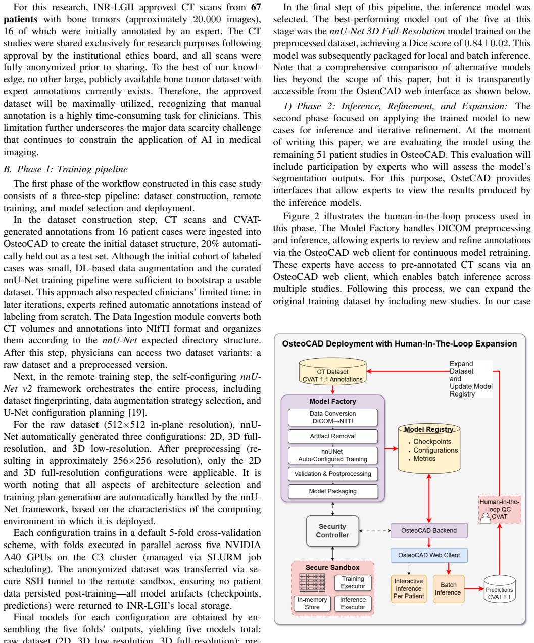
The load-bearing object is OsteoCAD's three-layer container architecture: a client-services layer that clinicians interact with through a web browser, an AI-services layer that preprocesses images and orchestrates training and inference, and an infrastructure layer that can run locally or connect through an encrypted tunnel to a remote GPU sandbox. The sandbox is deliberately ephemeral: anonymized data is decrypted only in volatile memory and purged when a task ends. Around that infrastructure, a human-in-the-loop loop is the mechanism that turns zero data into a usable dataset: clinicians correct machine-generated contours, and corrected cases feed the next training round. Training itself i
Load-bearing premise
The whole demonstration depends on the assumption that a Dice score of 0.84 measured on 16 annotated patients is representative of how the model will perform on the remaining 51 patients and in everyday clinical use.
What would settle it
Take the model trained on the 16-patient cohort and run it on all 51 remaining patient scans; report the per-case Dice distribution and compare against a second expert's manual contours. If the median is substantially below 0.84 or the spread is large, the claim that the framework produces clinically useful models in a no-data hospital does not yet stand.
If this is right
- A clinic starting with no labeled images can bootstrap a dataset by having experts correct machine predictions instead of labeling from scratch.
- Training that would require an in-house GPU cluster can be performed on remote ephemeral compute, with patient data kept in memory and purged after each task.
- The trained model and its performance metrics are presented in a web dashboard, so non-experts can select and deploy the best configuration.
- Because the framework is agnostic to the target label, the same infrastructure can serve different teams and different segmentation tasks on shared or separate datasets.
- Dataset expansion plus retraining is the intended operating loop: as the cohort grows from 16 to 67 patients, the deployed model is expected to improve.
Where Pith is reading between the lines
- The reported 0.84 Dice is best read as a proof-of-capability for the workflow rather than a benchmark for bone-tumor segmentation; the real test is whether per-case performance on the remaining 51 patients stays in the same range.
- A useful controlled experiment would measure annotation time saved by the human-in-the-loop loop relative to manual labeling from scratch; the paper asserts time savings but does not quantify them.
- If the 51-patient validation confirms the initial Dice score, the framework's contribution is a deployment recipe rather than a new segmentation algorithm, and it should be evaluated as such.
- The privacy-preserving remote-execution pattern could naturally extend to multi-center collaboration, letting each site keep data local while sharing only model-derived updates.
Editorial analysis
A structured set of objections, weighed in public.
Referee Report
Summary. The paper presents OsteoCAD, a modular three-layer container-based cloud–edge framework that integrates DICOM/CVAT ingestion, artifact removal, nnU-Net training, secure remote GPU execution via SSH, a model registry, and human-in-the-loop annotation for medical image segmentation. The authors validate the framework through a single-site case study at INR-LGII for large-bone tumor segmentation in CT: 67 patient studies were approved, 16 initially annotated, and the best of five nnU-Net configurations achieved a Dice score of 0.84±0.02. Phase 2, evaluation on the remaining 51 patients, is reported as in progress. The paper's central claim is that OsteoCAD can enable hospitals with minimal informatics support and no pre-existing annotated datasets to train, validate, and deploy state-of-the-art deep learning models in routine clinical workflows.
Significance. If the reported results hold, OsteoCAD would be a useful feasibility demonstration for bringing deep-learning segmentation to resource-constrained hospitals. The paper has concrete strengths: a real deployment at a rehabilitation center, use of established open components (nnU-Net, Niivue, CVAT), a modular design, attention to privacy and data sovereignty, and an explicit human-in-the-loop workflow. The authors also candidly state that phase-2 validation is incomplete. However, the quantitative evidence currently supporting the main claim is thin: one small-cohort result, an ambiguous evaluation protocol, post-hoc model selection, and no per-case or subgroup analysis. The architecture is plausible, but the evidence is not yet sufficient to support the strong 'routine clinical workflows' conclusion.
major comments (4)
- [§IV-B (Phase 1: Training pipeline)] The evaluation protocol is ambiguous and the effective test size is very small. The text states that '20% automatically held out as a test set' on the 16-patient cohort, and separately that each configuration uses nnU-Net's 'default 5-fold cross-validation scheme.' It is unclear whether the reported Dice 0.84±0.02 comes from the 20% holdout (about 3 patients) or from the mean over the five CV folds. If it is the holdout, the uncertainty estimate is not meaningful and no confidence interval is given; if it is the CV folds, the ±0.02 reflects fold variance, not uncertainty in the selected model. Please clarify the exact protocol, report per-patient Dice values, and provide confidence intervals.
- [§IV-B (model selection step)] The reported Dice is used to select 'the best-performing model out of the five' nnU-Net configurations. If the same evaluation set was used both to choose the model and to report its performance, the number is a maximum over five configurations and is positively biased. No selection correction, nested validation, or independent test result is provided. This is load-bearing because the entire feasibility claim rests on this single number. Please address the bias explicitly, for example by reporting all five models' Dice, using a true held-out test set untouched by selection, or presenting the phase-2 results on the 51 remaining patients as the unbiased estimate.
- [§IV-B1 and §V (Conclusions)] The central claim in Section V that OsteoCAD 'can enable hospitals ... to train, validate, and deploy state-of-the-art deep learning models in routine clinical workflows' goes beyond what the current data support. The paper itself states that evaluation on the remaining 51 patients is 'in progress' at the time of writing, so the only completed quantitative validation is the small, potentially selection-biased pilot. The conclusion should either be rephrased to 'initial feasibility' or, preferably, the revised paper should include the completed phase-2 evaluation with per-case results and subgroup or scanner-level analysis. Without this, the main contribution remains a promising prototype rather than a validated framework.
- [§V and §III-C] The paper repeatedly claims that OsteoCAD lowers technical barriers and can be used 'without demanding advanced technical expertise,' but no usability or workload evidence is provided. There is no measurement of setup time, annotation effort, training time, user training required, or a comparison to a manual nnU-Net deployment. For a systems/feasibility paper, reporting at least the clinician time for annotation and the total time from raw DICOM to deployed model would make this claim concrete. Without such data, the 'democratization' argument is asserted rather than demonstrated.
minor comments (6)
- [§IV-B1] The subsection is numbered '1) Phase 2' under 'Phase 1,' which is confusing; use a separate subsection number. Also, 'OsteCAD' is a typo for 'OsteoCAD.'
- [Throughout (§III-C, §IV-A)] 'CV AT' should be 'CVAT' (the annotation tool). The spacing appears in multiple places.
- [§IV-B] The list 'five models total: raw dataset (2D, 3D low-resolution, 3D full-resolution); preprocessed dataset (2D, 3D full-resolution)' is correct but could be clearer: the raw-variant low-resolution model is not evaluated on the preprocessed variant. A small table with each model's Dice and dataset variant would improve transparency.
- [§IV-A] The claim that 'no other large, publicly available bone tumor dataset with expert annotations currently exists' is strong and would benefit from a citation or a brief search statement.
- [§II (Related Work)] Reference [5] appears incomplete (missing full author list and venue). Please cite the published version of the nnU-Net revisited paper.
- [General] No code or data release is mentioned. For a framework paper, a repository link (even a private/demo version) would substantially aid reproducibility and allow reviewers to verify the claimed web-interface features.
Circularity Check
No equation-level circularity: the Dice result is an empirical measurement on held-out data; self-citations are peripheral integration components, not load-bearing reductions.
full rationale
The paper does not derive any target quantity from an input that already contains it. The central quantitative result, Dice 0.84±0.02, is presented in §IV-B as the performance of the nnU-Net 3D Full-Resolution model trained on the preprocessed dataset and evaluated against expert ground truth on a 20% held-out subset of the 16 initially annotated cases. The segmentation engine (nnU-Net, [19]) is external, and the metric is computed from actual predictions versus ground truth, not produced by an equation whose variables include the reported value. The framework claim in §V ('train, validate, and deploy state-of-the-art deep learning models') is supported by this case study and by the deployment architecture, not by a formal derivation. The two self-citations — [21] for artifact removal in the Data Ingestion module and [20] for secure data exchange — are cited as components of the pipeline, not as evidence that the Dice value must take a particular value; they do not carry the inference. The manuscript itself flags the main validation limitation: 'At the moment of writing this paper, we are evaluating the model using the remaining 51 patient studies in OsteoCAD' (§IV-B.1), and it states that a comprehensive model comparison 'lies beyond the scope of this paper' (§IV-B). These are acknowledged evidential gaps, not circular reasoning. One non-circular concern is that the reported Dice was obtained after choosing the 'best-performing model out of the five at this stage' on the same small held-out set, which can inflate the metric; this is a statistical-evaluation weakness (small n, post-hoc selection, no per-case distribution), not a case of a fitted input being renamed a prediction. Accordingly, the circularity burden is low: score 1 reflects the peripheral self-citations and the weak external anchoring of the single metric, without any identified reduction.
Axiom & Free-Parameter Ledger
axioms (4)
- domain assumption nnU-Net's automatic configuration produces a competitive segmentation model for this dataset without manual hyperparameter tuning.
- domain assumption Dice measured on the initial 16-patient cohort is representative of performance on the remaining 51 patients and of clinical utility.
- domain assumption The remote sandbox's in-memory processing, immediate memory purging, and SSH tunneling provide adequate data protection for real clinical use.
- domain assumption No other large publicly available expert-annotated bone-tumor CT dataset exists, justifying creation of a private dataset.
read the original abstract
Artificial Intelligence (AI) and Deep Learning (DL) have notably advanced medical image analysis, yet many health- care organizations struggle to adopt them due to limited com- putational resources and specialized expertise. To address these barriers, we introduce OsteoCAD, a modular eHealth framework that democratizes access to DL tools in clinical practice. Osteo- CAD delivers end-to-end DL capabilities-from dataset creation and preprocessing to model training and inference-through an integrated and user-friendly interface. To mitigate local hardware constraints, the framework securely connects to remote GPU infrastructures. We validate OsteoCAD's feasibility through a real-world case study in Mexico focused on large bone tumor segmentation. The results demonstrate the framework's ability to enable DL-powered eHealth solutions without demanding ad- vanced technical expertise or complex local configurations.
Figures



Reference graph
Works this paper leans on
-
[1]
A survey on deep learning in medical image analysis,
G. Litjens, T. Kooi, B. E. Bejnordi, A. A. A. Setio, F. Ciompi, M. Ghafoo- rian, J. A. van der Laak, B. van Ginneken, and C. I. S ´anchez, “A survey on deep learning in medical image analysis,”Medical Image Analysis, vol. 42, pp. 60–88, 12 2017
2017
-
[2]
Deep learning in medical imaging and radiation therapy,
B. Sahiner, A. Pezeshk, L. M. Hadjiiski, X. Wang, K. Drukker, K. H. Cha, R. M. Summers, and M. L. Giger, “Deep learning in medical imaging and radiation therapy,”Medical Physics, vol. 46, no. 1, pp. e1–e36, 1 2019
2019
-
[3]
AI-based computer-aided diagnosis (AI-CAD): the latest review to read first,
H. Fujita, “AI-based computer-aided diagnosis (AI-CAD): the latest review to read first,”Radiological Physics and Technology, vol. 13, no. 1, pp. 6–19, 3 2020. [Online]. Available: https://link.springer.com/article/10.1007/s12194-019-00552-4
-
[4]
U-net: Convolutional networks for biomedical image segmentation,
O. Ronneberger, P. Fischer, and T. Brox, “U-net: Convolutional networks for biomedical image segmentation,” inLecture Notes in Computer Science (including subseries Lecture Notes in Artificial Intelligence and Lecture Notes in Bioinformatics), vol. 9351. Springer Verlag, 2015, pp. 234–241
2015
-
[5]
nnU-Net Revisited: A Call for Rigorous Validation in 3D Medical Image Segmentation
F. Isensee, Tassilo Wald, C. Ulrich, M. Baumgartner, S. Roy, K. Maier- Hein, and P. F. J ¨ager, “nnU-Net Revisited: A Call for Rigorous Validation in 3D Medical Image Segmentation.” [Online]. Available: https://github.com/MIC-DKFZ/nnUNet
-
[6]
U-Net and Its Variants for Medical Image Segmentation: A Review of Theory and Applications,
N. Siddique, S. Paheding, C. P. Elkin, and V . Devabhaktuni, “U-Net and Its Variants for Medical Image Segmentation: A Review of Theory and Applications,”IEEE Access, vol. 9, pp. 82 031–82 057, 2021. [Online]. Available: https://ieeexplore.ieee.org/abstract/document/9446143
arXiv 2021
-
[7]
Barriers to and Facilitators of Artificial Intelligence Adoption in Health Care: Scoping Review
M. Hassan, A. Kushniruk, and E. Borycki, “Barriers to and Facilitators of Artificial Intelligence Adoption in Health Care: Scoping Review.”JMIR human factors, vol. 11, no. 1, p. e48633, 8 2024
2024
-
[8]
Improving Explainability and Integrability of Medical AI to Promote Health Care Professional Acceptance and Use: Mixed Systematic Review,
Y . Liu, C. Liu, J. Zheng, C. Xu, and D. Wang, “Improving Explainability and Integrability of Medical AI to Promote Health Care Professional Acceptance and Use: Mixed Systematic Review,”Journal of Medical Internet Research, vol. 27, no. 1, p. e73374, 8 2025. [Online]. Available: https://www.jmir.org/2025/1/e73374
2025
-
[9]
Artificial Intelligence Platform Architecture for Hospital Systems: Systematic Review Corresponding Author
M. Maimaitiaili, Y . Jiamaliding, G. Dai, H. Xiao, W. Kuerbanjiang, and Y . Yi, “Artificial Intelligence Platform Architecture for Hospital Systems: Systematic Review Corresponding Author.”
-
[10]
Radiologist shortage leaves patient care at risk, warns royal college,
Z. Kmietowicz, “Radiologist shortage leaves patient care at risk, warns royal college,” p. j3855, 8 2017
2017
-
[11]
3D Slicer as an image computing platform for the Quantitative Imaging Network,
A. Fedorov, R. Beichel, J. Kalpathy-Cramer, J. Finet, J. C. Fillion- Robin, S. Pujol, C. Bauer, D. Jennings, F. Fennessy, M. Sonka, J. Buatti, S. Aylward, J. V . Miller, S. Pieper, and R. Kikinis, “3D Slicer as an image computing platform for the Quantitative Imaging Network,”Magnetic Resonance Imaging, vol. 30, pp. 1323–1341, 2012
2012
-
[12]
I. Wolf, M. Vetter, I. Wegner, M. Nolden, T. Bottger, M. Hastenteufel, M. Schobinger, T. Kunert, and H.-P. Meinzer, “The medical imaging interaction toolkit (MITK): a toolkit facilitating the creation of interactive software by extending VTK and ITK,”https://doi.org/10.1117/12.535112, vol. 5367, pp. 16–27, 5 2004
-
[13]
ITK-SNAP: An interactive tool for semi-automatic segmentation of multi-modality biomedical images,
P. A. Yushkevich, Y . Gao, and G. Gerig, “ITK-SNAP: An interactive tool for semi-automatic segmentation of multi-modality biomedical images,” Proceedings of the Annual International Conference of the IEEE Engi- neering in Medicine and Biology Society, EMBS, vol. 2016-October, pp. 3342–3345, 10 2016
2016
-
[14]
Open Health Imaging Foundation Viewer: An Extensible Open-Source Framework for Building Web-Based Imaging Applications to Support Cancer Research,
E. Ziegler, T. Urban, D. Brown, J. Petts, S. D. Pieper, R. Lewis, C. Hafey, and G. J. Harris, “Open Health Imaging Foundation Viewer: An Extensible Open-Source Framework for Building Web-Based Imaging Applications to Support Cancer Research,”JCO Clinical Cancer Infor- matics, vol. 4, no. 4, p. CCI.19.00131
-
[15]
Niivue: a WebGL2 based medical image viewer supporting over 30 formats of volumes and meshes,
L. Overbeek, “Niivue: a WebGL2 based medical image viewer supporting over 30 formats of volumes and meshes,” https://github.com/niivue/niivue, 3 2021
2021
-
[16]
XNAT Central: Open Sourcing Imaging Research Data,
R. Herrick, W. Horton, T. Olsen, M. McKay, K. A. Archie, and D. S. Marcus, “XNAT Central: Open Sourcing Imaging Research Data,” NeuroImage, vol. 124, no. Pt B, p. 1093, 1 2015. [Online]. Available: https://pmc.ncbi.nlm.nih.gov/articles/PMC4965359/
2015
-
[17]
Parabricks: GPU Ac- celerated Universal Pan-Instrument Genomics Analysis Software Suite,
T. Zhu, P. Vats, S. Onken, A. Dunstan, B. Zamirai, D. F. Puleri, A. Nair, M. Oliva, A. Gaihre, P. Sadhnani, S. Li, K. Arumugam, A. Chacon, M. Maric, J. Cohen, A. Sethia, and M. Samadi, “Parabricks: GPU Ac- celerated Universal Pan-Instrument Genomics Analysis Software Suite,” bioRxiv, p. 2025.07.23.666378, 8 2025
2025
-
[18]
MONAI Label: A framework for AI-assisted interactive labeling of 3D medical images,
A. Diaz-Pinto, S. Alle, V . Nath, Y . Tang, A. Ihsani, M. Asad, F. P ´erez- Garc´ıa, P. Mehta, W. Li, M. Flores, H. R. Roth, T. Vercauteren, D. Xu, P. Dogra, S. Ourselin, A. Feng, and M. J. Cardoso, “MONAI Label: A framework for AI-assisted interactive labeling of 3D medical images,” Medical Image Analysis, vol. 95, no. 6, p. 103207, 7 2024
2024
-
[19]
nnU-Net: Self-adapting Framework for U-Net- Based Medical Image Segmentation,
F. Isensee, J. Petersen, A. Klein, D. Zimmerer, P. F. Jaeger, S. Kohl, J. Wasserthal, G. Koehler, T. Norajitra, S. Wirkert, and K. H. Maier-Hein, “nnU-Net: Self-adapting Framework for U-Net- Based Medical Image Segmentation,” 9 2018. [Online]. Available: http://arxiv.org/abs/1809.10486
Pith/arXiv arXiv 2018
-
[20]
Xook-Sec: A policy-as-code framework for secure data-sharing on the computing continuum: CA Torres-Charles et al
C. A. Torres-Charles, D. D. Sanchez-Gallegos, J. L. Gonzalez-Compean, M. Morales-Sandoval, and J. Carretero, “Xook-Sec: A policy-as-code framework for secure data-sharing on the computing continuum: CA Torres-Charles et al.”Cluster Computing, vol. 28, no. 14, p. 889, 2025
2025
-
[21]
A Computer-aided Framework for Detecting Osteosarcoma in Computed Tomography Scans,
M. Rodriguez-Herrero, D. D. Sanchez-Gallegos, M. A. N ´u˜nez-Gaona, H. Aguirre-Meneses, L. A. V . Guti´errez, M. I. G. Velasco, J. L. Gonzalez- Compean, and J. Carretero, “A Computer-aided Framework for Detecting Osteosarcoma in Computed Tomography Scans,” 3 2026
2026
discussion (0)
Sign in with ORCID, Apple, or X to comment. Anyone can read and Pith papers without signing in.