REVIEW 2 major objections 1 minor 2 cited by
The HECKTOR 2025 challenge benchmarks top algorithms at 0.75 Dice for PET/CT tumor segmentation, 0.66 C-index for survival prediction, and 0.56 balanced accuracy for HPV classification on a 10-center test set.
Reviewed by Pith at T0; open to challenge. T0 means a machine referee read the full paper against a public rubric. the ladder, T0–T4 →
T0 review · grok-4.3
2026-06-26 18:15 UTC pith:JO5MXEBR
load-bearing objection This is an incremental update to the HECKTOR challenge series with more data and an HPV task, but the reported scores rest on summary stats without the checks needed for clinical claims. the 2 major comments →
HEad and neCK TumOR (HECKTOR) 2025: Benchmark of Segmentation, Diagnosis, and Prognosis in Multimodal PET/CT
The pith
A machine-rendered reading of the paper's core claim, the machinery that carries it, and where it could break.
Core claim
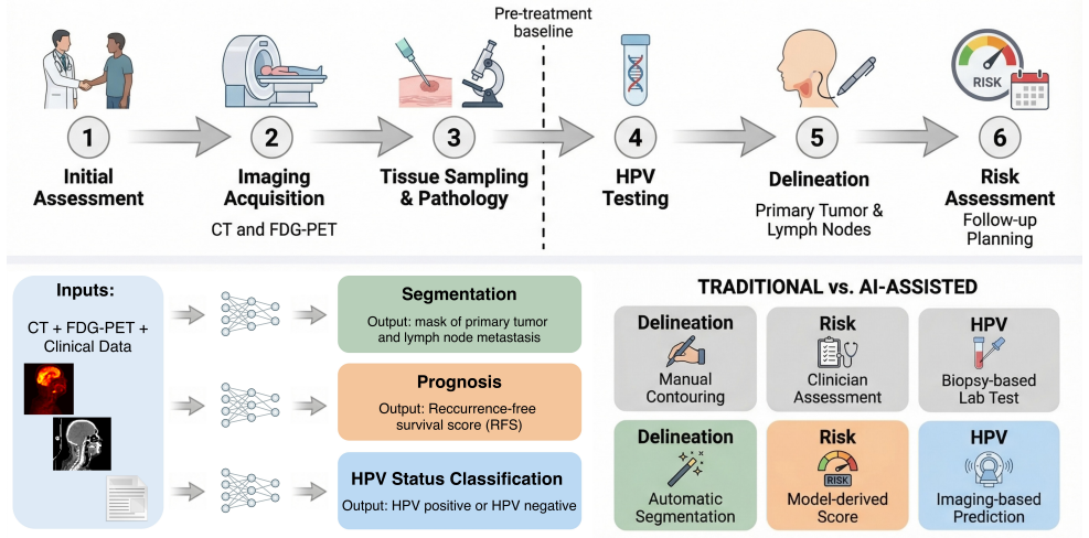
The HECKTOR 2025 challenge supplies an expanded multi-institutional PET/CT dataset and evaluates submitted methods on three tasks, with the strongest entries reaching a mean Dice similarity coefficient of 0.75 for gross tumor volume segmentation, a concordance index of 0.66 for recurrence-free survival prediction, and a balanced accuracy of 0.56 for noninvasive HPV status classification.
What carries the argument
The three-task benchmark on the held-out multi-center test set that measures segmentation accuracy, survival concordance, and diagnostic balance across lesion types.
Load-bearing premise
The held-out test set from the 10 centers is assumed to be representative of future clinical deployment distributions and that the chosen metrics directly translate to clinical utility without further validation on prospective or outcome-linked data.
What would settle it
A prospective multi-center trial that applies the top submitted methods to new patients and records whether the reported Dice, C-index, and accuracy values are reproduced or whether downstream clinical decisions change measurably.
If this is right
- Segmentation outputs at 0.75 Dice can reduce inter-observer variability and time in radiotherapy contouring.
- Survival models at 0.66 C-index supply a quantitative basis for risk grouping in treatment planning.
- HPV classifiers at 0.56 balanced accuracy offer an imaging-based complement to biopsy when tissue sampling is difficult.
- Performance differences across lesion characteristics highlight where current methods remain sensitive to tumor size or location.
- The public benchmark dataset enables direct comparison of new algorithms against these reference scores.
Where Pith is reading between the lines
- Methods that excel on one task may transfer to related oncology imaging problems if the same multi-center evaluation protocol is reused.
- Combining the three outputs into a single decision-support score could be tested by linking predictions to actual recurrence events in follow-up data.
- Extending the same data collection to additional imaging modalities or cancer sites would test whether the observed performance ceilings are modality-specific.
Editorial analysis
A structured set of objections, weighed in public.
Referee Report
Summary. The manuscript reports on the HECKTOR 2025 challenge, a multi-task benchmark for head and neck cancer analysis on multimodal PET/CT and EHR data from over 1,100 patients across 10 centers. It describes three tasks—segmentation of primary gross tumor volume (GTVp) and metastatic lymph nodes (GTVn), recurrence-free survival prediction, and HPV status classification—evaluated on a held-out test set with 15 final submissions from 35 registered teams. Top results are reported as mean Dice similarity coefficient of 0.75 for segmentation, concordance index of 0.66 for survival, and balanced accuracy of 0.56 for HPV classification, with analysis of methodologies and discussion of clinical implications.
Significance. If the performance numbers are supported by appropriate statistical validation, the work provides a useful standardized benchmark for multimodal HNC analysis that can facilitate method comparison and progress in automated radiotherapy planning and prognosis tools. The multi-center dataset spanning 10 institutions and the three complementary tasks represent a clear strength for the field.
major comments (2)
- [Abstract] Abstract: the reported aggregate metrics (mean Dice 0.75, C-index 0.66, balanced accuracy 0.56) are given as point estimates with no accompanying confidence intervals, p-values, or statistical testing details, which is load-bearing for the central performance claims on the held-out set.
- [Abstract] Abstract / Results: no lesion-specific breakdowns, inter-observer Dice reference, or baseline comparisons are referenced despite the claim of evaluating performance across lesion characteristics, limiting assessment of whether the top algorithms meaningfully exceed clinical variability.
minor comments (1)
- The abstract would be strengthened by stating the size of the held-out test set and the exact number of patients per task.
Simulated Author's Rebuttal
We thank the referee for the detailed review and constructive feedback on statistical reporting and clinical context. We address each major comment below.
read point-by-point responses
-
Referee: [Abstract] Abstract: the reported aggregate metrics (mean Dice 0.75, C-index 0.66, balanced accuracy 0.56) are given as point estimates with no accompanying confidence intervals, p-values, or statistical testing details, which is load-bearing for the central performance claims on the held-out set.
Authors: We agree that the abstract would benefit from explicit uncertainty quantification. The full manuscript reports bootstrapped 95% confidence intervals and pairwise statistical comparisons (Wilcoxon rank-sum tests with FDR correction) for the top submissions in the results section. We will revise the abstract to include the 95% CIs for the three headline metrics. revision: yes
-
Referee: [Abstract] Abstract / Results: no lesion-specific breakdowns, inter-observer Dice reference, or baseline comparisons are referenced despite the claim of evaluating performance across lesion characteristics, limiting assessment of whether the top algorithms meaningfully exceed clinical variability.
Authors: The results section already contains lesion-specific breakdowns (Dice stratified by GTVp vs. GTVn, primary tumor volume quartiles, and center) and places the observed Dice values in the context of published inter-observer variability for HNC (typically 0.65–0.82). Standard nnU-Net and 3D U-Net baselines are also reported. Because the abstract is space-constrained, we will add a single sentence summarizing that top methods reach but do not surpass literature inter-observer ranges and briefly note the lesion-stratified findings. revision: yes
Circularity Check
Empirical benchmark report with no derivations or self-referential predictions
full rationale
The paper is a challenge benchmark summary reporting Dice, C-index, and balanced accuracy on a held-out multi-center test set. No equations, fitted parameters, or first-principles derivations are present. No self-citation load-bearing steps, uniqueness theorems, or ansatzes are invoked. Central claims are direct empirical measurements, not reductions to inputs by construction. This matches the default non-circular case for benchmark papers.
Axiom & Free-Parameter Ledger
read the original abstract
Head and neck cancers (HNC) represent a significant global health burden, with accurate tumor delineation being essential for effective radiotherapy planning. The complexity of the oropharyngeal anatomy, combined with the heterogeneous appearance of tumors on imaging, makes manual segmentation time-intensive and subject to inter-observer variability. Beyond segmentation, predicting long-term clinical outcomes, such as recurrence-free survival (RFS), and determining human papillomavirus (HPV) status from noninvasive imaging, remain challenging yet clinically valuable goals. The HECKTOR 2025 challenge addresses these needs by establishing a comprehensive benchmark for automated HNC analysis using multimodal PET/CT imaging and electronic health records. Building on previous editions (2020-2022), this challenge features an expanded multi-institutional dataset comprising over 1,100 patients from 10 centers worldwide. Participants were tasked with three complementary objectives: (1) segmenting primary gross tumor volumes (GTVp) and metastatic lymph nodes (GTVn), (2) predicting recurrence-free survival, and (3) classifying HPV status. The challenge attracted 35 registered teams, with 15 final submissions evaluated on a held-out test set. Top-performing algorithms achieved a mean Dice similarity coefficient of 0.75 for segmentation, a concordance index of 0.66 for survival prediction, and a balanced accuracy of 0.56 for HPV classification. This paper presents a comprehensive analysis of the submitted methodologies, evaluates their performance across different lesion characteristics, and discusses their implications for clinical translation in automated oncology workflows and decision support systems.
Figures


Forward citations
Cited by 2 Pith papers
-
HERMES: A Hybrid Ensemble for Head-and-Neck Tumor Segmentation, TN Staging, and Recurrence-Free Survival on PET/CT
On HECKTOR 2026, mask-derived geometry features for nodal staging gave out-of-fold balanced accuracy 0.720 vs 0.691 for radiomics (paired CI includes zero); on ground-truth masks the gap was 0.897 vs 0.837.
-
Value-Monotonicity Matters: A Concordance Loss for Deep Survival Prediction
A sigmoid concordance loss whose value approximates one minus the C-index stays coupled to ranking performance throughout training, while likelihood-based survival losses provably and empirically decouple from it.
Reference graph
Works this paper leans on
-
[1]
Global burden of head and neck cancer: Epidemiological transitions, inequities, and projections to 2050,
H. Sun, M. Yu, Z. An, F. Liang, B. Sun, Y. Liu, and S. Zhang, “Global burden of head and neck cancer: Epidemiological transitions, inequities, and projections to 2050,”Frontiers in Oncology, vol. 15, p. 1665019, 2025
2050
-
[2]
Global burden of head and neck cancer from 1990 to 2021: A comprehensive analysis and projections to 2030 based on the global burden of disease study 2021,
M. Deng, Y. Lin, L. Yan, Z. Fei, C. Chen, and J. Ding, “Global burden of head and neck cancer from 1990 to 2021: A comprehensive analysis and projections to 2030 based on the global burden of disease study 2021,”PLOS One, vol. 20, 2025
1990
-
[3]
Global burden and future trends of head and neck cancer: a deep learning-based analysis (1980–2030),
Q. Hu, S. Lv, X. Wang, P. Pan, W. Gong, and J. Mei, “Global burden and future trends of head and neck cancer: a deep learning-based analysis (1980–2030),”PLOS One, vol. 20, 2025
1980
-
[4]
Head and neck cancer preven- tion: from primary prevention to impact of clin- icians on reducing burden
D. Hashim, E. Genden, M. Posner, M. Hashibe, and P. Boffetta, “Head and neck cancer preven- tion: from primary prevention to impact of clin- icians on reducing burden.”Annals of oncology : official journal of the European Society for Medi- cal Oncology, vol. 30 5, pp. 744–756, 2019
2019
-
[5]
Head and neck squamous cell carcinoma,
“Head and neck squamous cell carcinoma,”Nature Reviews Disease Primers, vol. 6, p. 1, 2020
2020
-
[6]
Antoch, N
G. Antoch, N. Saoudi, H. Kuehl, G. Dah- men, S. Mueller, T. Beyer, A. Bockisch, J. De- batin, and L. Freudenberg, “Accuracy of whole- body dual-modality fluorine-18-2-fluoro-2-deoxy- d-glucose positron emission tomography and com- puted tomography (fdg-pet/ct) for tumor staging in solid tumors: comparison with ct and pet.” Journal of clinical oncology : o...
2004
-
[7]
Automatic head and neck tumor segmentation and outcome prediction relying on fdg-pet/ct images: Findings from the second edi- tion of the hecktor challenge,
V. Andrearczyk, V. Oreiller, S. Boughdad, C. L. L. Rest, O. Tankyevych, H. Elhalawani, M. Jreige, J. O. Prior, M. Valli` eres, D. Visvikis, M. Hatt, and A. Depeursinge, “Automatic head and neck tumor segmentation and outcome prediction relying on fdg-pet/ct images: Findings from the second edi- tion of the hecktor challenge,”Medical image anal- ysis, vol....
2023
-
[8]
Results from the autopet challenge on fully au- tomated lesion segmentation in oncologic pet/ct imaging,
S. Gatidis, M. Fr¨ uh, M. Fabritius, and et al., “Results from the autopet challenge on fully au- tomated lesion segmentation in oncologic pet/ct imaging,”Nature Machine Intelligence, vol. 6, pp. 1396 – 1405, 2024
2024
-
[9]
Deep learning- based auto-delineation of gross tumour volumes and involved nodes in pet/ct images of head and neck cancer patients,
Y. M. Moe, A. R. Groendahl, O. Tomic, E. Dale, E. Malinen, and C. Futsaether, “Deep learning- based auto-delineation of gross tumour volumes and involved nodes in pet/ct images of head and neck cancer patients,”European Journal of Nu- clear Medicine and Molecular Imaging, vol. 48, pp. 2782 – 2792, 2021
2021
-
[10]
Characterization of pet/ct images using texture analysis: the past, the present. . . any future?
M. Hatt, F. Tixier, L. Pierce, P. Kinahan, C. L. L. Rest, and D. Visvikis, “Characterization of pet/ct images using texture analysis: the past, the present. . . any future?”European Journal of Nu- clear Medicine and Molecular Imaging, vol. 44, pp. 151–165, 2016
2016
-
[11]
Pre- dicting immunotherapy response in advanced solid tumors using quantitative imaging features from cd8 pet/ct exams
M. Postow, A. P. Peris, A. Estepa-Fern´ andez, A. F. Matanzo, A. J. Pastor, J. P. Fern´ andez, F. B. Bataller, K. Schmiedehausen, and M. Ferris, “Pre- dicting immunotherapy response in advanced solid tumors using quantitative imaging features from cd8 pet/ct exams.”Journal of Clinical Oncology, 2025
2025
-
[12]
Prediction of lung malignancy progression and survival with ma- chine learning based on pre-treatment fdg-pet/ct,
B. Huang, J. Sollee, Y. Luo, A. Reddy, Z. Zhong, J. Wu, J. Mammarappallil, T. Healey, G. Cheng, C. Azzoli, D. Korogodsky, P. J. Zhang, X. Feng, J. Li, Z. Jiao, and H. Bai, “Prediction of lung malignancy progression and survival with ma- chine learning based on pre-treatment fdg-pet/ct,” eBioMedicine, vol. 82, 2022
2022
-
[13]
Enhancing the con- touring efficiency for head and neck cancer radio- therapy using atlas-based auto-segmentation and scripting,
Y. Nagayasu, S. Ohira, T. Ikawa, A. Masaoka, N. Kanayama, T. Nishi, T. Kazunori, Y. Yoshino, 14 M. Miyazaki, Y. Uedaet al., “Enhancing the con- touring efficiency for head and neck cancer radio- therapy using atlas-based auto-segmentation and scripting,”in vivo, vol. 38, no. 4, pp. 1712–1718, 2024
2024
-
[14]
Head and neck tumor segmentation in pet/ct: The hecktor challenge,
V. Oreiller, V. Andrearczyk, M. Jreige, and et al., “Head and neck tumor segmentation in pet/ct: The hecktor challenge,”Medical image analysis, vol. 77, p. 102336, 2021
2021
-
[15]
Imaging biomarker roadmap for cancer studies,
“Imaging biomarker roadmap for cancer studies,” Nature reviews. Clinical oncology, vol. 14, pp. 169 – 186, 2016
2016
-
[16]
Biomarkers in head and neck squamous cell carcinoma: unravel- ing the path to precision immunotherapy,
K. S. Saini, S. Somara, H. C. Ko, P. Thatai, A. Quintana, Z. D. Wallen, M. F. Green, R. Mehro- tra, S. McGuigan, L. Panget al., “Biomarkers in head and neck squamous cell carcinoma: unravel- ing the path to precision immunotherapy,”Fron- tiers in Oncology, vol. 14, p. 1473706, 2024
2024
-
[17]
Predicting cancer outcomes with radiomics and artificial intelligence in radiology,
K. Bera, N. Braman, A. Gupta, V. Velcheti, and A. Madabhushi, “Predicting cancer outcomes with radiomics and artificial intelligence in radiology,” Nature Reviews Clinical Oncology, vol. 19, pp. 132– 146, 2021
2021
-
[18]
Prostate cancer radiogenomics—from imaging to molecular characterization,
M. Ferro, O. de Cobelli, M. Vartolomei, G. Lu- carelli, F. Crocetto, B. Barone, A. Sciarra, F. del Giudice, M. Muto, M. Maggi, G. Car- rieri, G. Busetto, U. Falagario, D. Terracciano, L. Cormio, G. Musi, and O. T˘ ataru, “Prostate cancer radiogenomics—from imaging to molecular characterization,”International Journal of Molec- ular Sciences, vol. 22, 2021
2021
-
[19]
Recent advances in deep learning and medical imaging for head and neck cancer treatment: Mri, ct, and pet scans,
M. Illimoottil and D. Ginat, “Recent advances in deep learning and medical imaging for head and neck cancer treatment: Mri, ct, and pet scans,” Cancers, vol. 15, 2023
2023
-
[20]
Benefits of automated gross tumor volume segmentation in head and neck cancer using multi-modality information
H. Bollen, S. Willems, M. Wegge, F. Maes, and S. Nuyts, “Benefits of automated gross tumor volume segmentation in head and neck cancer using multi-modality information.”Radiotherapy and oncology : journal of the European Society for Therapeutic Radiology and Oncology, p. 109574, 2023
2023
-
[21]
Artificial intelligence-based methods in head and neck cancer diagnosis: an overview,
H. Mahmood, M. Shaban, N. Rajpoot, and S. Khurram, “Artificial intelligence-based methods in head and neck cancer diagnosis: an overview,” British Journal of Cancer, vol. 124, pp. 1934 – 1940, 2021
1934
-
[22]
Global cancer observatory: Cancer to- day. lyon, france: International agency for research on cancer,
E. J, E. M, L. F, L. M, C. M, M. L, P. M, Z. A, S. I, and B. F, “Global cancer observatory: Cancer to- day. lyon, france: International agency for research on cancer,”World Health Organization, 2024
2024
-
[23]
A multicenter dataset for lymph node clinical target volume de- lineation of nasopharyngeal carcinoma,
X. Luo, W. Liao, Y. Zhaoet al., “A multicenter dataset for lymph node clinical target volume de- lineation of nasopharyngeal carcinoma,”Scientific Data, 2024
2024
-
[24]
A multimodal dataset for precision oncology in head and neck cancer,
M. D¨ orrich, M. Balk, T. Heusinger, S. Beyer, H. Kanso, C. Matek, A. Hartmann, H. Iro, M. Eck- stein, A.-O. Gostianet al., “A multimodal dataset for precision oncology in head and neck cancer,” medRxiv, pp. 2024–05, 2024
2024
-
[25]
Overview of the heck- tor challenge at miccai 2022: Automatic head and neck tumor segmentation and outcome prediction in pet/ct
A. V, O. V, A. M, and A. A, “Overview of the heck- tor challenge at miccai 2022: Automatic head and neck tumor segmentation and outcome prediction in pet/ct.”Head Neck Tumor Challenge, 2022
2022
-
[26]
N. Saeed, S. Hassan, S. Hardan, and et al., “A multimodal and multi-centric head and neck cancer dataset for segmentation, diagnosis and outcome prediction,” 2025. [Online]. Available: https://arxiv.org/abs/2509.00367
-
[27]
U-Net: Convolutional Networks for Biomedical Image Segmentation
O. Ronneberger, P. Fischer, and T. Brox, “U-net: Convolutional networks for biomedical image segmentation,” 2015. [Online]. Available: https://arxiv.org/abs/1505.04597
work page internal anchor Pith review Pith/arXiv arXiv 2015
-
[28]
Available: https://doi.org/10.1038/s41592-020-01008-z
F. Isensee, P. F. Jaeger, S. A. A. Kohl, J. Petersen, and K. H. Maier-Hein, “nnu-net: a self-configuring method for deep learning-based biomedical image segmentation,”Nature Methods, vol. 18, pp. 203–211, 2021. [Online]. Available: https://doi.org/10.1038/s41592-020-01008-z
-
[29]
A. Hatamizadeh, V. Nath, Y. Tang, D. Yang, H. Roth, and D. Xu, “Swin unetr: Swin transformers for semantic segmentation of brain tumors in mri images,” 2022. [Online]. Available: https://arxiv.org/abs/2201.01266
-
[30]
Unetr: Transformers for 3d medical image segmentation,
A. Hatamizadeh, Y. Tang, V. Nath, D. Yang, A. Myronenko, B. Landman, H. Roth, and D. Xu, “Unetr: Transformers for 3d medical image segmentation,” 2021. [Online]. Available: https://arxiv.org/abs/2103.10504
-
[31]
Deep learning techniques in pet/ct imaging: A comprehensive review from sinogram to image space,
M. Fallahpoor, S. Chakraborty, B. Pradhan, O. Faust, P. D. Barua, H. Chegeni, and R. Acharya, “Deep learning techniques in pet/ct imaging: A comprehensive review from sinogram to image space,”Computer Methods and Programs in Biomedicine, vol. 243, p. 107880, 2024. [Online]. Available: https://www.sciencedirect. com/science/article/pii/S0169260723005461
2024
-
[32]
Design and validate a dual-modality characteristic information fusion system based on probabilistic graphical models,
X. Xia, R. Zhang, X. Yao, G. Huang, and T. Tang, “Design and validate a dual-modality characteristic information fusion system based on probabilistic graphical models,”Research Square,
-
[33]
Available: https://doi.org/10
[Online]. Available: https://doi.org/10. 21203/rs.3.rs-2565336/v1
-
[34]
Fusion of medical imaging and electronic health records using deep learning: a systematic review and implementation guidelines,
S.-C. Huang, A. Pareek, S. Seyyedi, I. Banerjee, and M. Lungren, “Fusion of medical imaging and electronic health records using deep learning: a systematic review and implementation guidelines,” npj Digital Medicine, vol. 3, 12 2020
2020
-
[35]
Consistent estimation of the ex- pected brier score in general survival models with 15 right-censored event times
S. M. Gerds TA, “Consistent estimation of the ex- pected brier score in general survival models with 15 right-censored event times.”Biometrical Journal, 2006
2006
-
[36]
De- coding tumour phenotype by noninvasive imaging using a quantitative radiomics approach,
H. Aerts, E. Velazquez, and R. Leijenaar, “De- coding tumour phenotype by noninvasive imaging using a quantitative radiomics approach,”Nature Communications, 2014
2014
-
[37]
Katzman, Uri Shaham, Alexander Cloninger, Jonathan Bates, Tingting Jiang, and Yuval Kluger
J. L. Katzman, U. Shaham, A. Cloninger, J. Bates, T. Jiang, and Y. Kluger, “Deepsurv: personalized treatment recommender system using a cox proportional hazards deep neural network,” BMC Medical Research Methodology, vol. 18, no. 1, Feb. 2018. [Online]. Available: http: //dx.doi.org/10.1186/s12874-018-0482-1
-
[38]
Transformer-based deep survival analysis,
S. Hu, E. Fridgeirsson, G. v. Wingen, and M. Welling, “Transformer-based deep survival analysis,” inProceedings of AAAI Spring Symposium on Survival Prediction - Algo- rithms, Challenges, and Applications 2021, ser. Proceedings of Machine Learning Re- search, R. Greiner, N. Kumar, T. A. Gerds, and M. van der Schaar, Eds., vol. 146. PMLR, 2021, pp. 132–148...
2021
-
[39]
Summary from an in- ternational cancer seminar focused on human papillomavirus (hpv)-positive oropharynx can- cer, convened by scientists at iarc and nci,
A. R. Kreimer, A. K. Chaturvedi, L. Ale- many, and et al., “Summary from an in- ternational cancer seminar focused on human papillomavirus (hpv)-positive oropharynx can- cer, convened by scientists at iarc and nci,” Oral Oncology, vol. 108, p. 104736, 2020. [On- line]. Available: https://www.sciencedirect.com/ science/article/pii/S136883752030172X
2020
-
[40]
Human papillomavirus and survival of patients with oropharyngeal cancer
A. KK, H. J, W. R, W. R, R. DI, N.-T. PF, W. WH, C. CH, J. RC, L. C, K. H, A. R, S. CC, R. KP, and G. ML, “Human papillomavirus and survival of patients with oropharyngeal cancer.”N Engl J Med, 2010
2010
-
[41]
Radiomic features associated with hpv status on pretreatment computed tomography in oropharyn- geal squamous cell carcinoma inform clinical prog- nosis
S. B, Y. K, G. J, L. C, L. L, L. J, S. S, B. NM, K. CF, T. P, F. P, K. SA, L. J. Jr, and M. A, “Radiomic features associated with hpv status on pretreatment computed tomography in oropharyn- geal squamous cell carcinoma inform clinical prog- nosis.”Front Oncol., 2021
2021
-
[42]
Deep learning based hpv status prediction for oropha- ryngeal cancer patients
L. DM, P. JC, C. SE, W. JJ, and B. S., “Deep learning based hpv status prediction for oropha- ryngeal cancer patients.”Cancers (Basel)., 2021
2021
-
[43]
Pet/ct ra- diomics signature of human papilloma virus associ- ation in oropharyngeal squamous cell carcinoma
H. SP, M. A, Z. T, B. P, R. C, S. K, F. R, K. AS, K. BH, J. BL, P. ML, B. B, and P. S, “Pet/ct ra- diomics signature of human papilloma virus associ- ation in oropharyngeal squamous cell carcinoma.” Eur J Nucl Med Mol Imaging., 2020
2020
-
[44]
Less is more: Efficient PET/CT segmen- tation and multimodal prediction of recurrence- free survival and HPV status in head and neck can- cer,
L. Cai, X. Liang, T. Zhang, J. Huang, T. Tan, and Y. Yin, “Less is more: Efficient PET/CT segmen- tation and multimodal prediction of recurrence- free survival and HPV status in head and neck can- cer,” inFourth Head and Neck Cancer Tumor Le- sion Segmentation, Diagnosis and Prognosis, 2026
2026
-
[45]
Hec- tomixnet: Advancing automated head and neck tumor segmentation with multicenter PET/CT data,
M. Mazher, S. Niederer, and A. Qayyum, “Hec- tomixnet: Advancing automated head and neck tumor segmentation with multicenter PET/CT data,” inFourth Head and Neck Cancer Tumor Le- sion Segmentation, Diagnosis and Prognosis, 2026
2026
-
[46]
nnu-net v2 for head-and-neck PET/CT primary tumor and lymph node segmentation: A simple, strong baseline,
Y. Bu, Z. Wang, Y. Wang, J. Jiang, Y. Chen, and M. Lyu, “nnu-net v2 for head-and-neck PET/CT primary tumor and lymph node segmentation: A simple, strong baseline,” inFourth Head and Neck Cancer Tumor Lesion Segmentation, Diag- nosis and Prognosis, 2026
2026
-
[47]
Multi-stage multimodal progressive learning for coordinated segmentation, diagnosis, and progno- sis in head and neck cancer,
Y. Lin, S. Wu, H. Wang, L. Bi, and M. Meng, “Multi-stage multimodal progressive learning for coordinated segmentation, diagnosis, and progno- sis in head and neck cancer,” inFourth Head and Neck Cancer Tumor Lesion Segmentation, Diagno- sis and Prognosis, 2026
2026
-
[48]
A two-stage coarse-to-fine ensembling segmentation framework with multi- channel CT enhancement for head and neck tumor and lymph segmentation in PET and CT image,
C. Huang and L. Wang, “A two-stage coarse-to-fine ensembling segmentation framework with multi- channel CT enhancement for head and neck tumor and lymph segmentation in PET and CT image,” inFourth Head and Neck Cancer Tumor Lesion Segmentation, Diagnosis and Prognosis, 2026
2026
-
[49]
Enhancing survival outcomes in head and neck cancer through joint HPV classi- fication and tumor segmentation,
D. Chamseddine, S. Ahrari, M. Rizkallah, T. Car- lier, and D. Mateus, “Enhancing survival outcomes in head and neck cancer through joint HPV classi- fication and tumor segmentation,” inFourth Head and Neck Cancer Tumor Lesion Segmentation, Di- agnosis and Prognosis, 2026
2026
-
[50]
HECKTOR 2025 challenge: Hierarchi- cal multi-modal vision network for head-and-neck tumor segmentation and survival prediction,
B. Wu, E. Bao, Z. Li, Y. Chen, F. Qin, and Y. Chen, “HECKTOR 2025 challenge: Hierarchi- cal multi-modal vision network for head-and-neck tumor segmentation and survival prediction,” in Fourth Head and Neck Cancer Tumor Lesion Seg- mentation, Diagnosis and Prognosis, 2026
2025
-
[51]
Simplicity is all you need: out-of-the- box nnunet followed by binary-weighted radiomic model for segmentation and outcome prediction in head and neck pet/ct,
L. Rebaud, T. Escobar, F. Khalid, K. Girum, and I. Buvat, “Simplicity is all you need: out-of-the- box nnunet followed by binary-weighted radiomic model for segmentation and outcome prediction in head and neck pet/ct,” in3D head and neck tumor segmentation in PET/CT challenge. Springer, 2022, pp. 121–134
2022
-
[52]
A multi-modal deep learning framework for head and neck tumor seg- mentation and survival prediction,
F. Mao, Y. Jiang, N. Ji, Y. Jiang, X. Zheng, H. Zhang, and Y. Tang, “A multi-modal deep learning framework for head and neck tumor seg- mentation and survival prediction,” inFourth Head and Neck Cancer Tumor Lesion Segmentation, Di- agnosis and Prognosis, 2026
2026
-
[53]
From pixels to prognosis: Multimodal learning for head and neck cancer in the hecktor 2025 challenge,
B. Zhao and S. Ray, “From pixels to prognosis: Multimodal learning for head and neck cancer in the hecktor 2025 challenge,” inFourth Head and Neck Cancer Tumor Lesion Segmentation, Diag- nosis and Prognosis, 2026
2025
-
[54]
HECK- TOR2025 challenge report: Fully automated 16 diagnoses of HPV status using PET/CT images and clinical information,
M. Guo, T. Yu, J. He, and L. Xiang, “HECK- TOR2025 challenge report: Fully automated 16 diagnoses of HPV status using PET/CT images and clinical information,” inFourth Head and Neck Cancer Tumor Lesion Segmentation, Diag- nosis and Prognosis, 2026. [Online]. Available: https://openreview.net/forum?id=olICAFaTSD
2026
-
[55]
Multi-task deep learning for head and neck cancer: Segmentation, survival prediction, and HPV classification in the HECKTOR 2025 challenge,
J. Dexl and M. Ingrisch, “Multi-task deep learning for head and neck cancer: Segmentation, survival prediction, and HPV classification in the HECKTOR 2025 challenge,” inFourth Head and Neck Cancer Tumor Lesion Segmentation, Diagnosis and Prognosis, 2026. [Online]. Available: https://openreview.net/forum?id=YrJty0WWJn 17
2025
discussion (0)
Sign in with ORCID, Apple, or X to comment. Anyone can read and Pith papers without signing in.