REVIEW 4 major objections 4 minor 2 cited by
Unified Supervision For Vision-Language Modeling in 3D Computed Tomography
T0 review · 4 major / 4 minor · reviewed 2026-08-05 · deepseek-v4-flash
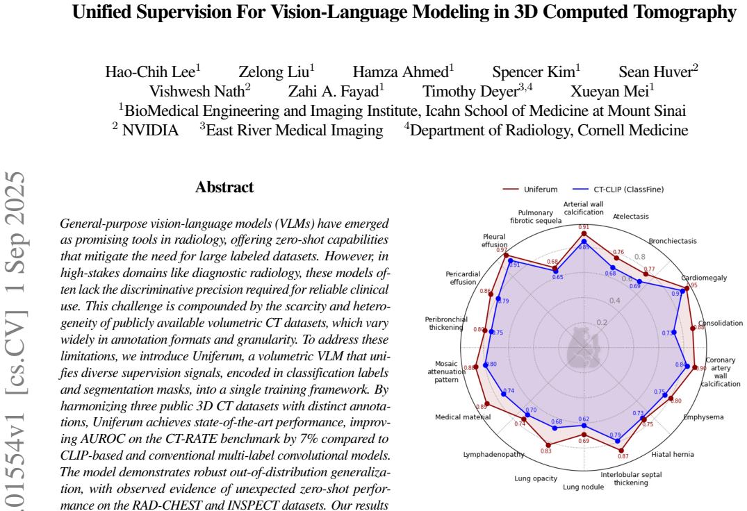
Pith's one-line read A vision-language model for 3D CT that trains on both diagnosis labels and organ masks, reaching 83.0% AUROC on CT-RATE (7 points above CLIP-based baselines) and showing unexpected zero-shot accuracy on withheld datasets.
desk verdict Useful empirical contribution on 3D CT VLMs, but the zero-shot generalization claim needs a clearer statement on data splitting and better label validation. read the letter →
The pith
A machine-rendered reading of the paper's core claim, the machinery that carries it, and where it could break.
The reading
What carries the argument
The task-conditioned fusion transformer: each training pair includes a natural-language task description, and the model predicts either a classification logit (from the CLS token) or a low-resolution patch-wise segmentation mask (from the per-patch embeddings) depending on which supervision the task supplies. This lets one backbone reuse every volume across many labels, mix classification and segmentation losses in a single batch, and transfer knowledge between datasets without requiring paired labels or masks.
What would settle it
Hold out segmentation entirely for the RAD-CHEST and INSPECT evaluation sets (do not run TotalSegmentator on those volumes, or remove their masks from segmentation training), then compare zero-shot AUROC. If the gains on unique labels drop to chance or shrink substantially, the zero-shot transfer claim is an artifact of domain adaptation to the segmentation outputs, not language-driven generalization.
Extended reading notes
Core claim
Uniferum reformulates every classification and segmentation label as a vision-language task: a CT volume is paired with a natural-language description such as 'Diagnose the presence of pleural effusion around the lungs' or 'Segment the lungs in the image,' and the model is trained to produce either a classification logit or a patch-wise segmentation prediction from the same fused embedding. A 3D EfficientNet encodes the volume; a small transformer fuses those features with the task text; the CLS token drives binary classification while per-patch embeddings predict downsampled masks. Because labels are decoupled, one volume participates in many tasks, and datasets with incompatible annotation
Load-bearing premise
The claim that adding segmentation masks is a neutral way to boost classification depends on the masks for the evaluation scans not leaking information; the reported zero-shot gains could instead come from the model having seen those same scans through their segmentation task.
Editorial extensions
If this is right
- If the claim holds, fragmented public CT datasets can be combined without re-annotation, making data-efficient 3D VLMs practical.
- Adding generic body segmentation (60 organ-level tasks) is a low-cost, universal way to improve classification AUROC by 1–3%.
- Task-conditioned evaluation lets a single model answer many diagnostic and prognostic questions, decoupling label count from model count.
- The observed zero-shot performance on unshared labels suggests language-conditioned representations generalize to unseen abnormalities, though this is the claim most sensitive to segmentation leakage.
Reading between the lines
- The segmentation gains may be anatomy-aware regularization rather than true multimodal reasoning; a cleaner ablation would hold out segmentation masks for the evaluation scans.
- The same task-conditioning trick could apply to other 3D modalities (MRI, PET) where annotations are similarly fragmented.
- The low-resolution mask objective (patch-wise u^3 prediction) may already provide enough spatial grounding, implying fine-grained voxel segmentation is unnecessary for classification gains.
- If zero-shot claims survive decontamination, they suggest that radiograph-level text prompts carry enough semantics to initialize unseen diagnostic categories, arguing for scaling task descriptions rather than datasets.
Editorial analysis
A structured set of objections, weighed in public.
Referee Report
Summary. The paper introduces Uniferum, a vision-language model for 3D CT that unifies classification labels and segmentation masks through task-conditioned training on CT-RATE, RAD-CHEST, and INSPECT. The model is an encoder-only transformer with a 3D EfficientNet vision encoder and text embeddings, trained with binary cross-entropy for classification and focal loss for segmentation. The authors report a 7% AUROC improvement over CT-CLIP on CT-RATE, zero-shot performance on INSPECT and RAD-CHEST unique tasks, and consistent 1–3% gains when body-segmentation tasks are added. The manuscript includes public code and comparisons with multiple baselines.
Significance. If the claims hold, the paper makes a useful contribution: it shows how heterogeneous public CT annotations can be harmonized in a single VLM, and it provides evidence that segmentation supervision can improve classification in a task-conditioned framework. The use of multiple datasets, public code, and some confidence-interval reporting are strengths. However, the central generalization and segmentation-benefit claims depend on experimental hygiene that is not fully described: the segmentation sampling may overlap with evaluation volumes, and some evaluation labels are machine-generated. These issues are fixable but need to be resolved before the conclusions can be accepted.
major comments (4)
- [§3.3, Table 2] The segmentation-task construction is described as 'randomly sampled an extra 10% samples from the dataset' without stating that this 10% is restricted to the training split. If INSPECT or RAD-CHEST validation/test volumes are included as segmentation targets, then the model has been trained on the exact volumes later used for 'zero-shot' evaluation. The +SEG improvements (e.g., INSPECT unique 0.5808→0.6130; RADCHEST unique 0.5559→0.5768; shared-task gains) could then reflect image-level domain adaptation rather than a transferable benefit of segmentation. This is load-bearing for the 'universal strategy' claim and the zero-shot claims. Please state explicitly that segmentation tasks are drawn only from training splits and, ideally, re-run the +SEG comparison with all evaluation volumes excluded from segmentation targets.
- [§3.4.3, §5.4] The 12 INSPECT diagnosis labels used for INSPECT-unique zero-shot evaluation are generated by keyword matching followed by GPT-4o-mini validation, with only 120 samples (10 per class) radiologist-verified (96% accuracy). The paper itself notes that labels such as 'nodules' may include false negatives. Because the zero-shot AUROC values are computed against these machine-generated labels, label noise could bias the reported comparisons. Please provide an analysis of label-noise sensitivity or restrict zero-shot evaluation to labels with stronger verification.
- [Table 2 and §5.2] The main AUROC table reports point estimates without confidence intervals or significance tests. This is particularly important for the 1–3% segmentation gains, which are small relative to likely variability, and for the headline 7% CT-RATE improvement. Some zero-shot examples include DeLong intervals, but the main claims and the 'consistently improved' statement rely on unquantified point estimates. Please add confidence intervals or significance tests for the key comparisons in Table 2.
- [§5.4] The p-values reported (e.g., p=0.03, p=0.02 for mortality improvements) are not corrected for multiple comparisons, and several confidence intervals overlap (e.g., honeycombing 69.2±11.0 vs. 65.0±13 after +SEG). The claim that body segmentation 'could be raised' by adding segmentation tasks is stated more strongly than the evidence supports. Please temper the wording or provide multiple-comparison-corrected statistics.
minor comments (4)
- [Conclusion] The conclusion states 'achieving 72% and 79% AUROC on out-of-distribution datasets RAD-CHEST and INSPECT respectively,' but Section 5.3 reports 72.1% on INSPECT shared and 79.6% on RAD-CHEST shared. The dataset names are swapped; please correct.
- [Throughout] There are several typographical and formatting issues (e.g., 'plumary emblosim', 'V alidation', 'W e', 'T o'). A careful proofread is needed.
- [§3.4.1] The CT-RATE validation description is slightly confusing: 'Scans of 100 patients from the official training set were withheld for model selection' and then the official validation set of 3002 volumes is used. Please clarify the relationship between the internal withheld set and the official validation set.
- [Table 2, baseline rows] The table includes dashes for some baseline cells. It would aid reproducibility to state in the caption which baseline results are from prior papers and which were produced by the authors.
Circularity Check
No significant circularity: the empirical claims are self-contained; a potential evaluation-contamination risk is noted but is not a derivation-level circularity.
full rationale
This paper is an empirical systems paper; its 'derivation chain' is a training/evaluation protocol rather than a mathematical derivation. The central claims—7% AUROC improvement on CT-RATE, out-of-distribution generalization, and zero-shot AUROC on INSPECT/RADCHEST unique labels—are produced by training a model on public datasets and evaluating on held-out label sets. I find no step where a reported quantity is defined in terms of another reported quantity, no parameter fitted to the target labels and then renamed as a prediction, and no load-bearing self-citation. The architecture (EfficientNet-b0 + transformer fusion) and losses are standard. The CT-CLIP/CT-NET baselines are cited from external work [6]. The body-segmentation masks come from TotalSegmentator, an external tool, and are used as auxiliary training targets; the classification labels used for evaluation are not used to fit the model. Thus the main empirical comparisons have independent content. One validity concern, not circularity: Section 3.3 says 'randomly sampled an extra 10% samples from the dataset' without specifying whether this 10% is split-restricted. If the segmentation tasks include INSPECT/RADCHEST validation or test volumes, the +SEG 'zero-shot' improvements in Section 5.4 could reflect image-level exposure rather than zero-shot transfer. This is a data-contamination risk that should be clarified, but it does not make the CT-RATE-only results circular, and it is not an equation-level reduction of a prediction to its inputs.
Assumptions & free parameters
free parameters (6)
- focal_loss_scale =
10
- negative_sampling_ratio =
1:1 for diagnostic tasks
- segmentation_extra_10% =
10% of samples per organ task
- LUNA16_upsample =
10x (6,010 samples)
- patch_size_u =
not specified
- transformer_layers =
4
assumptions (4)
- domain assumption TotalSegmentator organ masks are accurate enough to serve as training supervision.
- domain assumption GPT-4o-mini validated labels are accurate enough to serve as ground truth for 12 INSPECT diagnostic tasks.
- domain assumption Binary cross-entropy on text-conditioned tasks plus focal loss on masks jointly optimizes without negative transfer.
- domain assumption Task descriptions in natural language transfer across datasets, enabling zero-shot prediction.
Cite this review
Pith. "Pith review of Unified Supervision For Vision-Language Modeling in 3D Computed Tomography." pith.science (2026). https://pith.science/paper/ESM66PLM
@misc{pith2026250901554,
  author       = {Pith},
  title        = {Pith review of: Unified Supervision For Vision-Language Modeling in 3D Computed Tomography},
  year         = {2026},
  howpublished = {\url{https://pith.science/paper/ESM66PLM}},
  note         = {Machine review of arXiv:2509.01554}
}
read the original abstract
General-purpose vision-language models (VLMs) have emerged as promising tools in radiology, offering zero-shot capabilities that mitigate the need for large labeled datasets. However, in high-stakes domains like diagnostic radiology, these models often lack the discriminative precision required for reliable clinical use. This challenge is compounded by the scarcity and heterogeneity of publicly available volumetric CT datasets, which vary widely in annotation formats and granularity. To address these limitations, we introduce Uniferum, a volumetric VLM that unifies diverse supervision signals, encoded in classification labels and segmentation masks, into a single training framework. By harmonizing three public 3D CT datasets with distinct annotations, Uniferum achieves state-of-the-art performance, improving AUROC on the CT-RATE benchmark by 7% compared to CLIP-based and conventional multi-label convolutional models. The model demonstrates robust out-of-distribution generalization, with observed evidence of unexpected zero-shot performance on the RAD-CHEST and INSPECT datasets. Our results highlight the effectiveness of integrating heterogeneous annotations and body segmentation to enhance model performance, setting a new direction for clinically reliable, data-efficient VLMs in 3D medical imaging.
Figures


Figures from the paper (4 more)


Forward citations
Cited by 2 Pith papers
-
ORACLE-CT: Anatomy-Aware Support Pooling for CT Classification
ORACLE-CT improves CT classification performance by using anatomy-specific support pooling based on multi-organ segmentation, showing gains in AUROC on internal and external datasets.
-
Efficient Multimodal Clinical Question Answering for Pulmonary Embolism Risk Assessment
Benchmarks efficient MLLMs on eight PE QA tasks from the INSPECT dataset, finding stronger results with combined CTPA+EHR inputs and for diagnosis over prognosis.
Reference graph
Works this paper leans on
-
[1]
Fan Bai, Y uxin Du, Tiejun Huang, Max Q-H Meng, and Bo Zhao. M3d: Advancing 3d medical image analysis with multi-modal large language models.arXiv preprint arXiv:2404.00578, 2024
arXiv 2024
-
[2]
Louis Blankemeier, Joseph Paul Cohen, Ashwin Kumar, Dave V an V een, Syed Jamal Safdar Gardezi, Magdalini Paschali, Zhihong Chen, Jean-Benoit Delbrouck, Eduardo Reis, Cesar Truyts, et al. Merlin: A vision language foundation model for 3d computed tomography.arXiv preprint arXiv:2406.06512, 2024
arXiv 2024
-
[3]
3d-ct-gpt: Generating 3d radiology reports through integration of large vision-language models
Hao Chen, W ei Zhao, Yingli Li, Tianyang Zhong, Yisong W ang, Y oulan Shang, Lei Guo, Junwei Han, Tianming Liu, Jun Liu, et al. 3d-ct-gpt: Generating 3d radiology reports through integration of large vision-language models. arXiv preprint arXiv:2409.19330, 2024
arXiv 2024
-
[4]
Rachel Lea Draelos, David Dov, Maciej A Mazurowski, Joseph Y Lo, Ricardo Henao, Geoffrey D Rubin, and Lawrence Carin. Machine-learning-based multiple abnormality prediction with large-scale chest computed tomography volumes.Medical image analysis, 67:101857, 2021
work page 2021
-
[5]
Y u Gu, Robert Tinn, Hao Cheng, Michael Lucas, Naoto Usuyama, Xiaodong Liu, Tristan Naumann, Jianfeng Gao, and Hoifung Poon. Domain-specific language model pretraining for biomedical natural language processing.ACM Transactions on Computing for Healthcare (HEALTH), 3(1):1–23, 2021
work page 2021
-
[6]
Ibrahim Ethem Hamamci, Sezgin Er, Furkan Almas, A yse Gulni- han Simsek, Sevval Nil Esirgun, Irem Dogan, Muhammed Furkan Dasdelen, Omer Faruk Durugol, Bastian Wittmann, T amaz Amiranashvili, et al. Developing generalist foundation models from a multimodal dataset for 3d computed tomography.arXiv preprint arXiv:2403.17834, 2024
arXiv 2024
-
[7]
Xiaoyu He, Y ong W ang, Shuang Zhao, and Xiang Chen. Joint segmentation and classification of skin lesions via a multi-task learning convolutional neural network. Expert Systems with Applications, 230:120174, 2023
work page 2023
-
[8]
Shih-Cheng Huang, Liyue Shen, Matthew P Lungren, and Serena Y eung. Gloria: A multimodal global-local representation learning framework for label-efficient medical image recognition. In Proceedings of the IEEE/CVF international conference on computer vision, pages 3942–3951, 2021
work page 2021
Show all 31 references
-
[9]
Inspect: a multimodal dataset for patient outcome prediction of pulmonary embolisms.Advances in Neural Information Processing Systems, 36:17742–17772, 2023
Shih-Cheng Huang, Zepeng Huo, Ethan Steinberg, Chia-Chun Chiang, Curtis Langlotz, Matthew Lungren, Serena Y eung, Nigam Shah, and Jason Fries. Inspect: a multimodal dataset for patient outcome prediction of pulmonary embolisms.Advances in Neural Information Processing Systems,...
2023
-
[10]
Chexpert: A large chest radiograph dataset with uncertainty labels and expert comparison
Jeremy Irvin, Pranav Rajpurkar, Michael Ko, Yifan Y u, Silviana Ciurea-Ilcus, Chris Chute, Henrik Marklund, Behzad Haghgoo, Robyn Ball, Katie Shpanskaya, et al. Chexpert: A large chest radiograph dataset with uncertainty labels and expert comparison. In Proceedings of the AAAI...
2019
-
[11]
Radgraph: Extracting clinical entities and relations from radiology reports
Saahil Jain, Ashwin Agrawal, Adriel Saporta, Steven QH Truong, Du Nguyen Duong, T an Bui, Pierre Chambon, Y uhao Zhang, Matthew P Lungren, Andrew Y Ng, et al. Radgraph: Extracting clinical entities and relations from radiology reports. arXiv preprint arXiv:2106.14463, 2021
2021 arXiv
-
[12]
E3d-gpt: Enhanced 3d visual foundation for medical vision-language model.arXiv preprint arXiv:2410.14200, 2024
Haoran Lai, Zihang Jiang, Qingsong Y ao, Rongsheng W ang, Zhiyang He, Xiaodong T ao, W ei W ei, W eifu Lv, and S Kevin Zhou. E3d-gpt: Enhanced 3d visual foundation for medical vision-language model.arXiv preprint arXiv:2410.14200, 2024
2024 arXiv
-
[13]
T owards universal text-driven ct image segmentation
Y uheng Li, Y uxiang Lai, Maria Thor, Deborah Marshall, Zachary Buchwald, David S Y u, and Xiaofeng Y ang. T owards universal text-driven ct image segmentation. arXiv preprint arXiv:2503.06030, 2025
2025 arXiv
-
[14]
Focal loss for dense object detection
Tsung-Yi Lin, Priya Goyal, Ross Girshick, Kaiming He, and Piotr Doll´ar. Focal loss for dense object detection. InProceedings of the IEEE international conference on computer vision, pages 2980–2988, 2017
2017
-
[15]
Automatic joint segmentation and classification of breast ultrasound images via multi-task learning with object contextual attention.Frontiers in Oncology, 15:1567577, 2025
Y aling Lu, Fengyuan Sun, Jingyu W ang, and Kai Y u. Automatic joint segmentation and classification of breast ultrasound images via multi-task learning with object contextual attention.Frontiers in Oncology, 15:1567577, 2025
2025
-
[16]
Y -net: joint segmentation and classification for diagnosis of breast biopsy images
Sachin Mehta, Ezgi Mercan, Jamen Bartlett, Donald W eaver, Joann G Elmore, and Linda Shapiro. Y -net: joint segmentation and classification for diagnosis of breast biopsy images. In International conference on medical image computing and computer-assisted intervention, pages 8...
2018
-
[17]
A large-scale evaluation of automatic pulmonary nodule detection in chest ct using local image features and k-nearest-neighbour classification
Keelin Murphy, Bram van Ginneken, Arnold MR Schilham, BJ De Hoop, Hester A Gietema, and Mathias Prokop. A large-scale evaluation of automatic pulmonary nodule detection in chest ct using local image features and k-nearest-neighbour classification. Medical image analysis, 13(5)...
2009
-
[18]
Learning transferable visual models from natural language supervision
Alec Radford, Jong W ook Kim, Chris Hallacy, Aditya Ramesh, Gabriel Goh, Sandhini Agarwal, Girish Sastry, Amanda Askell, Pamela Mishkin, Jack Clark, et al. Learning transferable visual models from natural language supervision. In International conference on machine learning, p...
2021
-
[19]
3d convolutional neural networks for stalled brain capillary detection
Roman Solovyev, Alexandr A Kalinin, and T atiana Gabruseva. 3d convolutional neural networks for stalled brain capillary detection. Computers in biology and medicine, 141:105089, 2022
2022
-
[20]
Efficientnet: Rethinking model scaling for convolutional neural networks
Mingxing T an and Quoc Le. Efficientnet: Rethinking model scaling for convolutional neural networks. In International conference on machine learning, pages 6105–6114. PMLR, 2019
2019
-
[21]
Medclip: Contrastive learning from unpaired medical images and text
Zifeng W ang, Zhenbang Wu, Dinesh Agarwal, and Jimeng Sun. Medclip: Contrastive learning from unpaired medical images and text. In Proceedings of the Conference on Empirical Methods in Natural Language Processing. Conference on Empirical Methods in Natural Language Processing,...
2022
-
[22]
T otalseg- mentator: robust segmentation of 104 anatomic structures in ct images
Jakob W asserthal, Hanns-Christian Breit, Manfred T Meyer, Maurice Pradella, Daniel Hinck, Alexander W Sauter, T obias Heye, Daniel T Boll, Joshy Cyriac, Shan Y ang, et al. T otalseg- mentator: robust segmentation of 104 anatomic structures in ct images. Radiology: Artificial ...
2023
-
[23]
Navigating data scarcity using foundation models: A benchmark of few-shot and zero-shot learning approaches in medical imaging
Stefano W oerner and Christian F Baumgartner. Navigating data scarcity using foundation models: A benchmark of few-shot and zero-shot learning approaches in medical imaging. In International W orkshop on F oundation Models for General Medical AI, pages 30–39. Springer, 2024
2024
-
[24]
T owards generalist foundation model for radiology
Chaoyi Wu, Xiaoman Zhang, Y a Zhang, Y anfeng W ang, and W eidi Xie. T owards generalist foundation model for radiology. arXiv preprint arXiv:2308.02463, 2023
2023 arXiv
-
[25]
Learning a multi-task transformer via unified and customized instruction tuning for chest radiograph interpretation
Lijian Xu, Ziyu Ni, Xinglong Liu, Xiaosong W ang, Hongsheng Li, and Shaoting Zhang. Learning a multi-task transformer via unified and customized instruction tuning for chest radiograph interpretation. arXiv preprint arXiv:2311.01092, 2023
2023 arXiv
-
[26]
A large language model for electronic health records.NPJ digital medicine, 5(1): 194, 2022
Xi Y ang, Aokun Chen, Nima PourNejatian, Hoo Chang Shin, Kaleb E Smith, Christopher Parisien, Colin Compas, Cheryl Martin, Anthony B Costa, Mona G Flores, et al. A large language model for electronic health records.NPJ digital medicine, 5(1): 194, 2022
2022
-
[27]
Evaluating progress in automatic chest x-ray radiology report generation
Feiyang Y u, Mark Endo, Rayan Krishnan, Ian Pan, Andy Tsai, Eduardo Pontes Reis, Eduardo Kaiser Ururahy Nunes Fonseca, Henrique Min Ho Lee, Zahra Shakeri Hossein Abad, Andrew Y Ng, et al. Evaluating progress in automatic chest x-ray radiology report generation. P atterns, 4(9), 2023
2023
-
[28]
Umit: Unifying medical imaging tasks via vision-language models
Haiyang Y u, Siyang Yi, Ke Niu, Minghan Zhuo, and Bin Li. Umit: Unifying medical imaging tasks via vision-language models. arXiv preprint arXiv:2503.15892, 2025
2025 arXiv
-
[29]
Biomedclip: a multimodal biomedical foundation model pretrained from fifteen million scientific image-text pairs
Sheng Zhang, Y anbo Xu, Naoto Usuyama, Hanwen Xu, Jaspreet Bagga, Robert Tinn, Sam Preston, Rajesh Rao, Mu W ei, Naveen V alluri, et al. Biomedclip: a multimodal biomedical foundation model pretrained from fifteen million scientific image-text pairs. arXiv preprint arXiv:2303....
2023 arXiv
-
[30]
Knowledge-enhanced visual-language pre-training on chest radiology images
Xiaoman Zhang, Chaoyi Wu, Y a Zhang, W eidi Xie, and Y anfeng W ang. Knowledge-enhanced visual-language pre-training on chest radiology images. Nature Communications, 14(1):4542, 2023
2023
-
[31]
Mediclip: Adapting clip for few-shot medical image anomaly detection
Ximiao Zhang, Min Xu, Dehui Qiu, Ruixin Y an, Ning Lang, and Xiuzhuang Zhou. Mediclip: Adapting clip for few-shot medical image anomaly detection. In International Conference on Medical Image Computing and Computer-Assisted Intervention, pages 458–468. Springer, 2024. Unified ...
2024
Reviewed August 5, 2026 · model on record in the stance chip above.
Discussion (0). Sign in to comment.