REVIEW 3 major objections 4 minor 5 references
The paper claims that approximating 3D vertebra volumes from optimized 2D projections is a viable proxy for full 3D segmentation in cervical spine fracture recognition, reaching vertebra-level F1 of 68.15 and patient-level F1 of 82.26 on th
Reviewed by Pith at T0; open to challenge. T0 means a machine referee read the full paper against a public rubric. the ladder, T0–T4 →
T0 review · deepseek-v4-flash
2026-08-03 08:54 UTC pith:SOD4NN3J
load-bearing objection A thorough engineering pipeline paper whose central claim—that 2D-projection masks can replace full 3D segmentation for fracture recognition—is never actually tested in a controlled same-classifier comparison. the 3 major comments →
Tracing 3D Anatomy in 2D Strokes: A Multi-Stage Projection Driven Approach to Cervical Spine Fracture Identification
The pith
A machine-rendered reading of the paper's core claim, the machinery that carries it, and where it could break.
Core claim
The paper's central claim is that projection-based mask approximation is a viable proxy for full 3D vertebra segmentation in cervical fracture recognition: segmenting vertebrae from optimized 2D sagittal and coronal projections, back-projecting the masks into approximate 3D volumes, and classifying those volumes yields fracture-detection performance comparable to a full 3D-segmentation baseline. The evidence is the reported vertebra-level F1 of 68.15 and patient-level F1 of 82.26 under 5-fold cross-validation on the full imbalanced public training set, within about one F1 point of the reproduced single-model winner's 69.74 and 82.56. The authors interpret this as showing that the downstream
What carries the argument
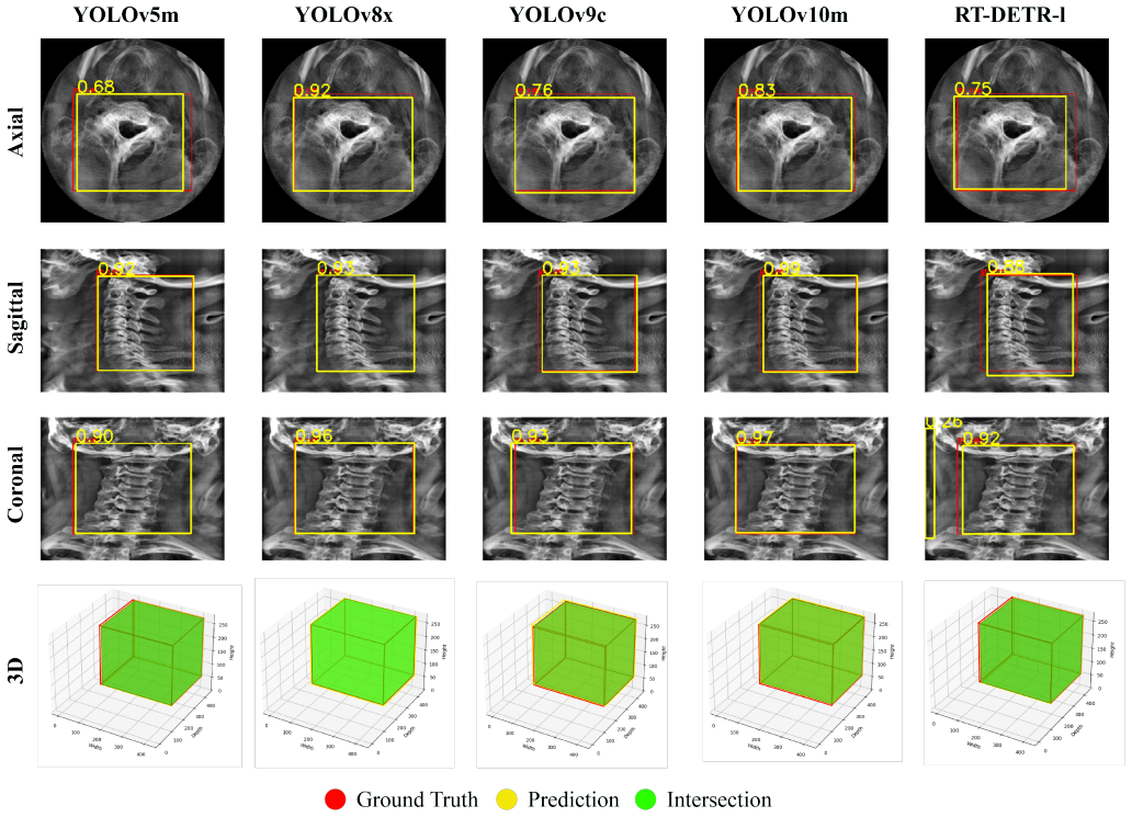
The load-bearing mechanism is approximate 3D vertebra mask construction from 2D projections: a detector localizes the cervical spine on variance projections, then an encoder-decoder network performs multi-label segmentation (each projected pixel may belong to several vertebrae because overlapping anatomy) on energy-based sagittal and coronal projections. The predicted 2D masks are extruded along their viewing axes and intersected to form approximate 3D masks, which crop vertebra volumes. Those volumes feed an ensemble of two 2.5D spatio-sequential CNN-Transformer models—one processing 15 evenly spaced slice stacks, the other processing 15 maximum-intensity-projection stacks—whose scores are
Load-bearing premise
The load-bearing premise is that comparing the projection pipeline to a reproduced single-model winner with a different classifier architecture (3D segmentation plus CNN-LSTM) fairly isolates the effect of using 2D-projection masks; because the paper never runs its own classifier on ground-truth 3D masks, a same-classifier comparison could reveal a larger gap than the reported one-point difference.
What would settle it
Train the identical 2.5D ensemble on vertebra volumes cropped with the ground-truth 3D masks (for the patients where masks exist, or an extended set) and compare vertebra-level F1 under the same 5-fold protocol. If the ground-truth-cropped classifier substantially outperforms the projection-cropped version—say, by more than the noise of cross-validation—then the projection approximation, not the classifier, is the bottleneck and the central claim fails.
If this is right
- Full 3D vertebra segmentation is not an absolute prerequisite for high-accuracy cervical fracture classification; 2D-derived volumes can carry the diagnostic signal.
- Localization and segmentation stages can run in lower-dimensional projection space, reducing the volumetric preprocessing burden relative to direct 3D methods.
- Fusing a slice-stack model with a maximum-intensity-projection-stack model improves vertebra-level precision and F1 over either model alone (score fusion: F1 68.15 vs 66.54 for the slice-stack model).
- The adaptive patient-level aggregation—a lower threshold when the two models agree—gives a balanced sensitivity of 82.52% and specificity of 83.55%, which the paper argues is more clinically useful than the high-precision, low-sensitivity 'if-any' rule.
- The model's saliency maps align with annotated fracture locations, and its agreement with the reference standard on a 30-case subset (vertebra-level κ = 0.711) is comparable to or above the three expert radiologists, supporting use as a second reader.
Where Pith is reading between the lines
- The paper never trains its own 2.5D classifier on volumes cropped with ground-truth 3D masks; such an ablation would directly measure how much diagnostic context the 2D-projection approximation loses, independent of architecture differences. I would expect this to be the decisive test of the proxy claim.
- If the projection proxy holds at scale, the same trick—reduce a 3D segmentation problem to two orthogonal 2D segmentations and fuse by intersection—may transfer to other anatomies where structures overlap in projection views, such as the thoracolumbar spine or rib cage, where volumetric segmentation is expensive.
- The multi-label formulation (allowing one projected pixel to belong to multiple vertebrae) is a general answer to a common problem: any projection of overlapping 3D anatomy creates class overlap, so multi-label supervision may be a reusable ingredient beyond vertebrae.
- A testable extension suggested by the paper's own limitations: replacing the fixed 15 equally spaced slices with anatomy-aware slice selection, and combining several projection operators as inputs, could recover part of the information lost in the 2D bottleneck.
Editorial analysis
A structured set of objections, weighed in public.
Referee Report
Summary. The paper proposes a three-stage pipeline for cervical spine fracture detection from CT: (1) YOLOv8-based localization of the cervical spine ROI using variance projections, (2) DenseNet121-UNet multi-label segmentation of C1–C7 on energy projections, and (3) reconstruction of approximate 3D vertebra masks by intersecting back-projected 2D masks, followed by an ensemble of 2.5D spatio-sequential CNN-Transformer classifiers. On the full RSNA 2022 training set the authors report vertebra-level F1 68.15 and patient-level F1 82.26, and they conclude from a comparison with a reproduced single-model RSNA winner that projection-based mask approximation is a viable proxy for full 3D vertebra segmentation in cervical fracture recognition.
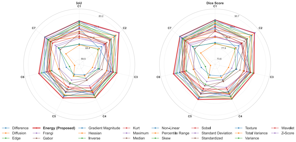
Significance. If the central claim were directly supported, the paper would make a meaningful contribution: it would show that computationally expensive full 3D segmentation is not required for this downstream classification task, while still achieving competitive performance on a large, imbalanced, clinically realistic dataset. The paper has several strengths: evaluation on the full RSNA 2022 training set with 5-fold cross-validation, a systematic comparison of 23 projection techniques, an interobserver variability study involving three radiologists, and public preprocessing/inference code. These elements increase the potential value of the work. However, the key causal claim—that the projection-derived masks, rather than other components of the pipeline, are responsible for the observed performance—is not tested by the experiments as presented.
major comments (3)
- [Section 4.3.6 / Table 9] The central claim—that projection-derived masks are a viable proxy for full 3D segmentation—is not directly tested. The comparison in Table 9 is between the full projection pipeline (YOLOv8 + projection segmentation + 2.5D ensemble) and a reproduced single-model RSNA winner (3D segmentation + CNN-LSTM). This changes the mask source, the classifier architecture, and training details simultaneously. A controlled experiment using the same 2.5D classifier trained on vertebra crops from ground-truth 3D masks versus crops from the estimated projection masks is needed to isolate the effect of mask approximation. Without that, the abstract's assertion that 'full 3D segmentation is not an absolute prerequisite' is not supported.
- [Abstract and Table 7] There is an inconsistency in the reported patient-level AUC. The abstract states 'area under the receiver operating characteristic curve of 91.62 and 90.95' in one version and '91.62 and 83.04' in the full-text abstract. Table 7 shows AUC = 90.95 for the 'If Any' fusion row and AUC = 83.04 for the 'Adaptive Avg' row. Since the abstract pairs the adaptive-threshold F1 (82.26) with a patient AUC, it should be paired with 83.04, not 90.95. Please correct the abstract and ensure all reported metrics are aligned with the table.
- [Section 4.2.1 and 4.2.2] The authors assert that 'small boundary inaccuracies are unlikely to meaningfully affect downstream vertebra volume cropping' (Section 4.2.1) and that the downstream task 'relies on accurate vertebra centered cropping rather than exact subvoxel boundary placement' (Section 4.2.2). This is an assumption, not a demonstrated result. The controlled comparison in major comment #1 would directly test this assumption; until such evidence is provided, the statement should be phrased as a hypothesis rather than a conclusion.
minor comments (4)
- [Section 4.3.6] The text says 'Our ensemble model achieved a vertebra-level F1-score of 66.54% and 93.54% accuracy,' but these values correspond to the non-fusion 'Proposed Method' in Table 9; the fusion model ('Proposed (Fusion)') achieves 68.15% and 94.51%. Please clarify which model is being referred to.
- [Section 3.3.1] There are two subsections numbered 3.3.1: 'Bounding box generation from 3D mask data' and 'Sequential Slice Selection.' Renumber the latter (e.g., 3.3.2) and adjust subsequent numbering.
- [Section 4.3.6] The estimated median vertebra-level F1 of approximately 67.57% 'from the organizer-provided vertebra-level F1 box plots' should be clearly labeled as an approximate reading of published figures, and the specific figure source should be cited.
- [Abstract] Two versions of the abstract are present (the metadata abstract and the full-text abstract) with different patient-level AUC values (90.95 vs. 83.04). Ensure the metadata abstract is synchronized with the manuscript.
Circularity Check
No circular derivation; the fracture labels, segmentation supervision, and external RSNA-winner baseline are independent of the fitted outputs.
full rationale
The paper's central claim is that projection-derived masks are a viable proxy for full 3D segmentation in fracture recognition. The derivation chain is not circular: fracture labels come from the independent RSNA reference standard ('Vertebra-wise fracture labels from the RSNA dataset were utilized to perform fracture classification'), and the segmentation model is trained on 87 ground-truth masks under 5-fold cross-validation, not on the fracture labels or the downstream classifier's predictions. The predicted masks are then used to crop vertebra volumes, and the classifier is trained on those crops with fracture labels. There is no equation in which a reported prediction is defined as the fitted parameter or in which the input already contains the output. The comparison to the RSNA 1st-place solution uses an external, publicly released single-model implementation ('we evaluated the publicly released single model implementation of the first-place solution using the same cross-validation protocol'), so it is an external benchmark rather than a self-citation chain. The paper's own limitation section candidly states that multi-stage design creates error-propagation risk and that only 87 patients have ground-truth masks, which is a validity caveat, not circularity. The skeptic's objection—that the baseline differs in architecture and training, and that no same-classifier GT-mask-crop control is run—is a real threat to the causal interpretation of the proxy claim, but it is a confounding/control problem, not a definitional or self-referential reduction of the result to its inputs. No circular step can be quoted from the paper, so the circularity score is 0.
Axiom & Free-Parameter Ledger
free parameters (5)
- Bounding-box tolerance t =
20 pixels
- Sagittal slice selection range =
100-420
- Positive-class loss weight w =
2
- Stack parameters for 2.5D model =
15 stacks, 5 slices per stack, 75 sampled slices
- Adaptive patient-level threshold =
not specified numerically
axioms (5)
- domain assumption RSNA fracture labels are ground truth for training and evaluation
- domain assumption The 87 patients with segmentation masks are representative of all 2,019 patients
- domain assumption Energy projection preserves vertebral boundaries and variance projection preserves spine localization
- domain assumption Intersection of extruded sagittal and coronal masks is an adequate 3D vertebra approximation
- standard math Cubic spline interpolation to at least 400 slices preserves diagnostic information
read the original abstract
Cervical spine fractures require rapid and accurate diagnosis, yet automatic CT interpretation remains challenging as subtle injuries must be assessed across large 3D volumes. We ask whether full 3D vertebra segmentation is necessary for automated fracture recognition, or whether vertebra masks approximated from 2D projections can preserve sufficient diagnostic context. We propose an end-to-end pipeline that localizes the cervical spine, estimates C1-C7 vertebra masks from optimized 2D projections, and uses the resulting vertebra-level volumes for downstream fracture classification. A YOLOv8 detector first localizes spine regions of interest from multi-view variance projections, achieving a 3D mean Intersection over Union of 94.45%. Multi-label vertebra segmentation is then performed with a DenseNet121-Unet on energy-based sagittal and coronal projections, attaining a mean Dice score of 87.86%. The predicted 2D masks are back-projected and fused into approximate 3D masks for each vertebra to extract volumes of interest from the original CT. These volumes are analyzed by an ensemble of 2.5D spatio-sequential CNN-Transformer models, yielding vertebra-level and patient-level F1 scores of 68.15 and 82.26, area under the receiver operating characteristic curve of 91.62 and 90.95, and area under the precision-recall curve of 75.60 and 92.00, respectively. The projection-derived volumes achieved fracture-recognition performance comparable to a full 3D-segmentation baseline, while shifting the vertebra segmentation stage into a lower-dimensional domain. Saliency-based explainability and interobserver variability analysis further examine interpretability and reliability. Overall, the results indicate that projection-based mask approximation is a viable proxy for full 3D vertebra segmentation in cervical fracture recognition.
Figures





Reference graph
Works this paper leans on
-
[3]
arXiv preprint arXiv:2405.14458
Yolov10: Real-time end-to-end object detection. arXiv preprint arXiv:2405.14458 . Wiegreffe, S., Pinter, Y ., 2019. Attention is not not explanation. arXiv preprint arXiv:1908.04626 . Wu, Z., Xia, G., Zhang, X., Zhou, F., Ling, J., Ni, X., Li, Y .,
Pith/arXiv arXiv 2019
-
[5]
Cervical spine fracture detection and classification using two-stage deep learning methodology. IEEE Access . Zanza, C., Tornatore, G., Naturale, C., Longhitano, Y ., Sa- viano, A., Piccioni, A., Maiese, A., Ferrara, M., V olonnino, G., Bertozzi, G., 2023. Cervical spine injury: Clinical and medico-legal overview. La radiologia medica 128, 103–112. Zhang,...
arXiv 2023
-
[2022]
Computers in Biology and Medicine 151, 106190
A novel 3d lumbar vertebrae location and segmentation method based on the fusion envelope of 2d hybrid visual projection images. Computers in Biology and Medicine 151, 106190. Yaseen, M., Ali, M., Ali, S., Hussain, A., Joo, M., Kim, H.C.,
-
[2023]
Physics in Medicine & Biology 68, 115010
Cervical spine fracture detection in computed tomogra- phy using convolutional neural networks. Physics in Medicine & Biology 68, 115010. Goodfellow, I., Bengio, Y ., Courville, A., 2016. Section 5.5: Estimators, Bias, and Variance. MIT Press. book section 5. Guha Paul, S., Saha, A., Assaduzzaman, M., 2023. A real-time deep learning approach for classifyi...
arXiv 2016
-
[2024]
Australian Journal of Multi-Disciplinary Engineering , 1–9
Identification of cervical spine fracture using deep learn- ing. Australian Journal of Multi-Disciplinary Engineering , 1–9. Golla, A.K., Lorenz, C., Buerger, C., Lossau, T., Klinder, T., Mutze, S., Arndt, H., Spohn, F., Mittmann, M., Goelz, L.,
discussion (0)
Sign in with ORCID, Apple, or X to comment. Anyone can read and Pith papers without signing in.