REVIEW 3 major objections 6 minor 47 references
Nanopillar-Driven Antibacterial Surfaces: Elucidating Bactericidal Mechanisms and Engineering Nanostructures for Enhanced Efficacy
T0 review · 3 major / 6 minor · reviewed 2026-08-10 · deepseek-v4-flash
Pith's one-line read Nanopillar bactericidal action splits into tip-tearing and contact-piercing depending on membrane stiffness.
desk verdict Interesting full-cell CG result on tip-adjacent tearing, but the two-mechanism story confounds bending rigidity with loading rate and needs the missing control before it can be believed. read the letter →
The pith
A machine-rendered reading of the paper's core claim, the machinery that carries it, and where it could break.
The reading
What carries the argument
The machinery is a one-particle-thick coarse-grained membrane whose pair potential has a repulsive-attractive radial part and an angular part that penalizes misorientation, giving parameters that tune the membrane's bending rigidity. The rigidity is matched to real bacterial membranes through the height-fluctuation spectrum $\langle |h(q)|^2 \rangle = k_B T/(K_c q^4)$, and an extra body force called the loading rate supplies the adhesive, gravitational, and hydrodynamic driving that the Lennard-Jones pillar-membrane term only partially captures. This lets the model simulate whole 500-nm spherical and 1800-nm cylindrical bacteria rather than membrane patches, and it is what allows failure to nucleate at a specific point of contact.
What would settle it
Look for the first breach: a low-rigidity membrane on 200-nm-tall, 170-nm-spaced pillars should tear at the pillar tip, not in the sagged span between pillars. A three-layer membrane simulation or time-resolved imaging of the same geometry could settle whether the rupture site matches the model.
Extended reading notes
Core claim
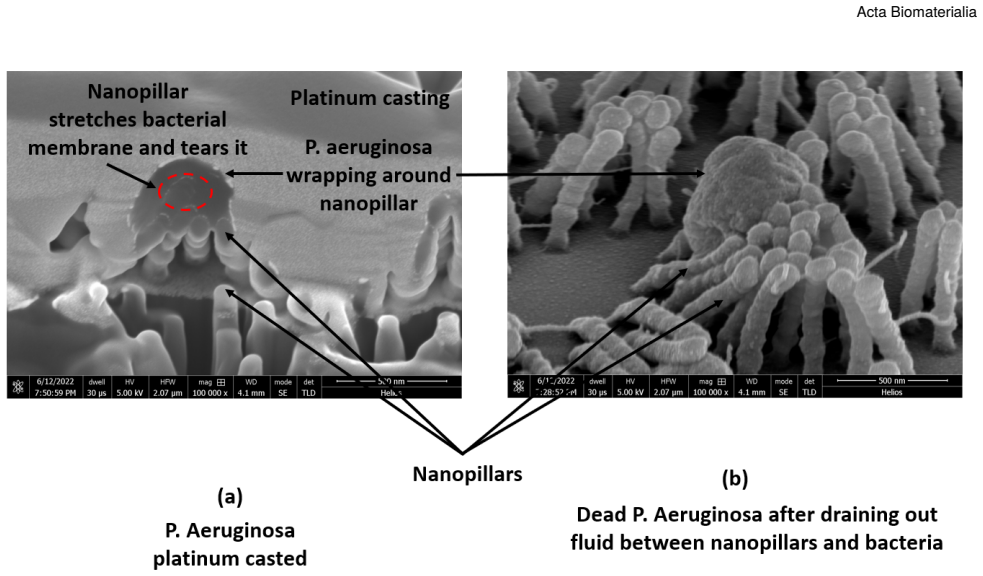
The central claim is that bacterial membranes on nanopillar arrays fail by one of two mechanisms controlled by membrane bending rigidity. For low-rigidity membranes, calibrated to gram-negative values near 15 $k_BT$, the membrane deforms locally at the pillar contact, slides and sags, and tears close to the nanopillar tip when the pillar is tall enough to suspend the bacterium; this contradicts the earlier picture that rupture occurs in the sagged region between the pillars. For high-rigidity membranes, calibrated to gram-positive values near 45 $k_BT$, the bacterium rests on the pillar tops with little deformation and survives, and only a roughly threefold increase in the imposed loading rate switches the failure mode to piercing at the contact point. Nanopillar height above a bacterium-dependent threshold and spacing below a value set by the bacterium's width are the design parameters that determine whether the tearing mode activates.
Load-bearing premise
The whole mechanism rests on the assumption that a one-particle-thick membrane tuned only to match bending rigidity, driven by an added loading-rate force, fails the same way and at the same location as a real three-layer bacterial envelope during roughly 30 minutes of contact.
Editorial extensions
If this is right
- Surface designers can use pillar height and spacing as the controls that switch a nanopillared surface from harmless to bactericidal for flexible-walled cells.
- For stiff-walled cells, geometry alone is not enough: the simulations require about a threefold higher loading rate to activate the piercing mechanism.
- When pillar spacing exceeds the width of the target bacterium, cells slip between pillars and survive, so spacing must stay below that width for reliable killing.
- Small spherical bacteria can squeeze into wider gaps more easily than large rod-shaped ones, so dense pillar arrays are needed for them.
Reading between the lines
- Beyond the paper, the mechanism map implies a stiffness-selective design space: a surface could kill gram-negative pathogens while sparing gram-positive cells, a prediction testable in mixed-culture viability assays.
- The nanosecond-scale simulation is bridged to the 30-minute contact process only by the adjustable loading rate; converting that rate into measured approach velocities and adhesion energies would make the model quantitative under realistic flow and settling conditions.
- Because the claimed failure site for flexible membranes is the pillar tip, independently varying tip curvature while holding height and spacing fixed would directly test the mechanism against the older sagging-between-pillars picture.
- In quiet, low-impact fluid conditions the model implies that piercing is rare, so nanopillar coatings under static incubation should be inherently more lethal to gram-negative than gram-positive bacteria, which could be checked with flow-versus-static experiments.
Signed reviews
Editorial analysis
A structured set of objections, weighed in public.
Referee Report
Summary. This manuscript combines experimental viability assays on polyimide nanopillar arrays with coarse-grained molecular dynamics simulations to study how nanopillar height, spacing, and bacterial membrane bending rigidity determine bactericidal efficacy. The simulations use a single-layer, one-particle-thick membrane model calibrated to reproduce literature values of bending rigidity via Helfrich-spectrum analysis, and a uniform body force called the loading rate is applied to the bacterium to mimic unaccounted attractive interactions. The authors report two distinct failure mechanisms: low bending rigidity membranes, representing gram-negative bacteria, tear near the nanopillar tip, while high bending rigidity membranes, representing gram-positive bacteria, are pierced at the point of contact when the loading rate is increased. They also report that increasing nanopillar height enhances bactericidal activity beyond a critical threshold and that increasing spacing reduces it; these trends are qualitatively consistent with their own experiments on E. coli/P. aeruginosa and S. aureus.
Significance. The paper introduces a computationally efficient coarse-grained membrane model that captures bacterial membrane bending rigidity, a key mechanical property in mechano-bactericidal studies, and it demonstrates in LAMMPS that membrane response to nanopillars depends on rigidity, pillar height, and spacing. The qualitative predictions—that gram-negative (low-rigidity) membranes tear near the pillar tip and are more easily killed than gram-positive (high-rigidity) membranes, and that taller pillars are more bactericidal—are consistent with the authors' own experiments, which is a genuine strength. If the mechanism can be confirmed with proper controls, it would refine the biophysical picture beyond the commonly cited sagging-between-pillars mechanism and would offer design guidance for nanopillar height and spacing. The model is implemented in a standard public MD code and the bending-rigidity calibration procedure is described in enough detail to be reproduced.
major comments (3)
- [§5 (Bactericidal mechanisms) and Table 1] The central claim that low bending rigidity causes tip-adjacent tearing while high bending rigidity causes contact piercing is confounded with the loading rate. In Table 1, the low-rigidity tearing cases (rows 1-3) are run at L.R. = 0.0002 eV/(m·Å), while the high-rigidity piercing case (row 7) is run at L.R. = 0.001, a five-fold increase. No low-rigidity membrane is simulated at L.R. = 0.001, and no high-rigidity membrane is simulated at L.R. = 0.0002 for longer times, so the two mechanisms could be selected by the loading rate rather than by bending rigidity. The Adhesion section (Fig. 13) further shows that changing the LJ epsilon from 0.02 to 2 or 50 switches failure from tearing to piercing, adding another uncontrolled parameter. A two-factor matrix of bending rigidity and loading rate, with at least the four corner cases, is required to attribute the mechanism to bending rigidity.
- [§3 (Coarse-Grained Model for Outer Fluid and Cytoplasm) and §5] The loading rate is an uncalibrated body force whose physical meaning is not established. The manuscript states that 'we use an additional force on bacteria i.e loading rate to simulate the attractive forces not accounted for using the LJ interactions,' and the membrane is modeled as a single-layer thin elastic layer with structural details neglected. The magnitude of 0.0002 eV/(m·Å) is not derived from estimates of gravity, adhesion, or hydrodynamic forces, and the paper does not justify why a membrane failure at ~1 ns corresponds to bactericidal outcomes at 30 minutes. Because the two claimed mechanisms are activated by changing this free parameter (and by changing epsilon), the simulated tearing and piercing cannot yet be confidently identified with the physical mechanisms of bactericidal action.
- [§5 (Figs. 14 and 15)] The claim of a threshold bending rigidity near 33.2 KbT separating tearing from survival is based on a single simulation per condition. There are no replicate runs, error bars, or sensitivity analysis for the CG model parameters. Given that the literature values themselves carry uncertainty (13±5 KbT for gram-negative, 43±5 KbT for gram-positive membranes), the threshold is within the combined uncertainty and is not statistically supported. This undermines the quantitative comparison between low- and high-rigidity bacteria.
minor comments (6)
- [Table 1] Table 1 lists the low-rigidity cases with B.R. = 8.2 KbT, but the text defines gram-negative membranes as 15.1 KbT; the units of L.R. in the table ('eV. mole)/(Å.grams)') are also inconsistent with the text's 'eV/(m·Å)'.
- [Abstract and Conclusion] The abstract and conclusion state that a threefold increase in loading rate is required for piercing, but Table 1 shows 0.001/0.0002 = 5, so the quantitative claim should be corrected.
- [Introduction] The name 'Xinelei et al. (9)' appears to refer to Li and Chen (ref. 9); the citation should be fixed.
- [Fig. 20 caption] The Fig. 20 caption contains a typo: 'his suggests' should be 'This suggests.'
- [References] Several references are incomplete: ref. 2 lacks journal and volume, and ref. 22 lacks publication details.
- [Adhesion section] The statement that ΔG_iwi = -0.0093 mJ/m^2 is 'very low compared to' -0.00963 mJ/m^2 is confusing, as the two values are nearly identical; rephrase.
Circularity Check
Piercing mechanism is demonstrated only at a chosen 5x higher loading rate; the claimed rigidity dependence of the piercing mode is partly forced by the input, while the tearing and height/spacing trends are genuine simulation outputs.
-
fitted input called prediction
[Abstract; Table 1, Case 7; 'Bactericidal mechanisms' section (Fig. 17)]
"A threefold increase in the loading rate is required for the piercing mechanism to activate and effectively kill gram-positive bacteria. ... For killing a gram-positive bacterium we need to increase the loading rate, as shown in Fig. 17, where the piercing mechanism activates."
The only piercing simulation is Table 1, Case 7: h=2000, B.R.=45.4, L.R.=0.001. All low-rigidity 'tearing' cases and all surviving high-rigidity cases use L.R.=0.0002, and no low-rigidity/high-LR control is reported. The conclusion that high bending rigidity 'requires' the higher loading rate therefore restates the input value chosen for the high-rigidity piercing run rather than a measured threshold. The paper's own description of LR as an ad hoc compensation force ('we use an additional force on bacteria i.e loading rate to simulate the attractive forces not accounted for using the LJ interactions') reinforces that the mechanism-selection result is partly by construction.
full rationale
The model is calibrated against independent literature values for membrane bending rigidity (13±5 and 43±5 kBT), and the tearing-vs-survival behavior across pillar height and spacing follows from the MD trajectories rather than being encoded directly in the potential, so most of the paper is not circular. The genuine circular/forced element is the piercing claim: the only high-rigidity piercing simulation uses L.R.=0.001, a 5x increase over the 0.0002 used in all low-rigidity and surviving high-rigidity cases, and no low-rigidity/high-LR control is reported. The conclusion that high bending rigidity 'requires' the higher loading rate therefore restates the chosen input value rather than a measured threshold; the 'threefold increase' in the abstract is not even the 5x ratio in Table 1. The ad hoc nature of LR is acknowledged by the authors ('we use an additional force on bacteria i.e loading rate to simulate the attractive forces not accounted for using the LJ interactions'), which makes the mechanism-selection result partly by construction. Self-citations (refs. 22-25, 36) support computational methodology and material parameters but are not load-bearing for the central mechanism; ref. 36 is used for polyimide contact angle and SEM interpretation, not to forbid alternatives.
Assumptions & free parameters
free parameters (5)
- Bending rigidity values =
15.1, 24.8, 33.2, 40.2, 45.4 kBT
- Loading rate =
0.0002 eV/(m*Å) per CG atom; 0.001 for piercing
- LJ epsilon for membrane-nanopillar interaction =
0.02 (baseline)
- CG fluid membrane potential parameters =
e.g., 0.2, 103, 200, 4, 5, 0 for low rigidity (order unclear)
- Simulation duration =
0.5 to 1.0 ns
assumptions (5)
- domain assumption The bacterial membrane is a thin elastic layer characterized solely by bending rigidity; internal structure neglected.
- ad hoc to paper A uniform additional force (loading rate) captures the net effect of gravity, adhesion, and hydrodynamics.
- domain assumption Real gram-negative and gram-positive membrane bending rigidities are 13±5 kBT and 43±5 kBT, respectively.
- standard math Helfrich relation <|h(q)|^2> = kBT/(Kc q^4) applies to this CG membrane.
- ad hoc to paper Observed 1 ns simulation outcomes correspond to 30-minute bactericidal outcomes.
Cite this review
Pith. "Pith review of Nanopillar-Driven Antibacterial Surfaces: Elucidating Bactericidal Mechanisms and Engineering Nanostructures for Enhanced Efficacy." pith.science (2026). https://pith.science/paper/QTYLEY7E
@misc{pith2026250111727,
  author       = {Pith},
  title        = {Pith review of: Nanopillar-Driven Antibacterial Surfaces: Elucidating Bactericidal Mechanisms and Engineering Nanostructures for Enhanced Efficacy},
  year         = {2026},
  howpublished = {\url{https://pith.science/paper/QTYLEY7E}},
  note         = {Machine review of arXiv:2501.11727}
}
read the original abstract
Insects like dragonflies and cicadas possess nanoprotusions on their wings that rupture bacterial membranes upon contact, inspiring synthetic antibacterial surfaces mimicking this phenomenon. Designing such biomimetic surfaces requires understanding the mechanical interaction between nanopillars and bacterial membranes. However, the small scales of these interactions pose challenges. Molecular Dynamics simulations offer precise and efficient modeling at these scales. This study presents a coarse-grained membrane model to explore the mechanical responses of gram-positive and gram-negative bacterial membranes to nanopillar arrays. By varying bacterial shapes (spherical and cylindrical), membrane bending rigidity, and loading rates, we identified two distinct failure mechanisms. Low bending rigidity, typical of gram-negative bacteria, leads to tearing near nanopillar tips, contrary to prior assumptions. High bending rigidity, characteristic of gram-positive bacteria, results in puncturing at contact points. Gram-positive bacteria are more resistant, requiring a threefold increase in loading rate for effective piercing. Nanopillar height and spacing also critically impact bactericidal efficacy: greater heights enhance activity beyond a critical threshold, while increased spacing reduces efficacy. This simplified coarse-grained model, representing bacterial membranes with high fidelity, enables cost-effective, full-scale simulations over extended periods. Our findings provide essential insights for optimizing nanopillared surface designs, advancing antibacterial technology through tailored height and spacing configurations.
Figures


Figures from the paper (16 more)




Reference graph
Works this paper leans on
-
[1]
Ivanova, E. P., J. Hasan, H. K. Webb, V. K. Truong, G. S. Watson, J. A. Watson, V. A. Baulin, S. Pogodin, J. Y. Wang, M. J. Tobin, C. Löbbe, and R. J. Crawford,
-
[2]
Ivanova, E. P., J. Hasan, H. K. Webb, G. Gervinskas, S. Juodkazis, V. K. Truong, A. H. Wu, R. N. Lamb, V. A. Baulin, G. S. Watson, J. A. Watson, D. E. Mainwaring, and R. J. Crawford. Bactericidal activity of black silicon
-
[3]
Hasan, J., H. K. Webb, V. K. Truong, S. Pogodin, V. A. Baulin, G. S. Watson, J. A. Watson, R. J. Crawford, and E. P. Ivanova, 2013. Selective bactericidal activ- ity of nanopatterned superhydrophobic cicada Psaltoda claripennis wing surfaces.Appl. Microbiol. Biotechnol. 97:9257–9262
work page 2013
-
[4]
Diu, T., N. Faruqui, T. Sjöström, B. Lamarre, H. F. Jenk- inson, B. Su, and M. G. Ryadnov, 2014. Cicada-inspired cell-instructive nanopatterned arrays.Scientific Reports 4:7122
work page 2014
-
[5]
Sjöström, T., A. H. Nobbs, and B. Su, 2016. Bactericidal nanospike surfaces via thermal oxidation of Ti alloy substrates. Materials Letters167:22–26
work page 2016
-
[6]
Hazell, G., P. W. May, P. Taylor, A. H. Nobbs, C. C. Welch, and B. Su, 2018. Studies of black silicon and black diamond as materials for antibacterial surfaces. Biomater. Sci.6:1424–1432
work page 2018
-
[7]
Pogodin, S., J. Hasan, V. A. Baulin, H. K. Webb, V. K. Truong, T. H. Phong Nguyen, V. Boshkovikj, C. J. Fluke, G. S. Watson, J. A. Watson, R. J. Crawford, and E. P. Ivanova, 2013. Biophysical model of bacterial cell inter- actionswithnanopatternedcicadawingsurfaces. Biophys. J. 104:835–840. 22 Manuscript submitted to Acta Biomaterialia Acta Biomaterialia
work page 2013
-
[8]
Xue, F., J. Liu, L. Guo, L. Zhang, and Q. Li, 2015. Theo- retical study on the bactericidal nature of nanopatterned surfaces. Journal of Theoretical Biology385:1–7
work page 2015
Show all 47 references
-
[9]
Enhancementandsuppression effects of a nanopatterned surface on bacterial adhesion
Li,X.,andT.Chen,2016. Enhancementandsuppression effects of a nanopatterned surface on bacterial adhesion. Phys. Rev. E 93:052419. https://link.aps.org/ doi/10.1103/PhysRevE.93.052419
2016 doi
-
[10]
Advanced Materials Interfaces6:1801646
Watson,G.S.,D.W.Green,J.A.Watson,Z.Zhou,X.Li, G.S.P.Cheung,andM.Gellender,2019.ASimpleModel for Binding and Rupture of Bacterial Cells on Nanopillar Surfaces. Advanced Materials Interfaces6:1801646
2019
-
[11]
Boseman, A
Nowlin, K., A. Boseman, A. Covell, and D. LaJe- unesse, 2015. Adhesion-dependent rupturing of Sac- charomyces cerevisiae on biological antimicrobial nanos- tructuredsurfaces. JournalofTheRoyalSocietyInterface 12:20140999
2015
-
[12]
Bandara, C. D., S. Singh, I. O. Afara, A. Wolff, T. Tes- famichael, K. Ostrikov, and A. Oloyede, 2017. Bacte- ricidal effects of natural nanotopography of dragonfly wing on Escherichia coli.ACS Appl. Mater. Interfaces 9:6746–6760
2017
-
[13]
Hazell, G., L. E. Fisher, W. A. Murray, A. H. Nobbs, and B. Su, 2018. Bioinspired bactericidal surfaces with poly- mer nanocone arrays.J. Colloid Interface Sci.528:389– 399
2018
-
[14]
Silhavy, T. J., D. Kahne, and S. Walker, 2010. The bacterialcellenvelope. ColdSpringHarb.Perspect.Biol. 2:a000414
2010
-
[15]
Kelleher, S. M., O. Habimana, J. Lawler, B. O’ Reilly, S. Daniels, E. Casey, and A. Cowley, 2016. Cicada wing surfacetopography:Aninvestigationintothebactericidal properties of nanostructural features.ACS Appl. Mater. Interfaces8:14966–14974
2016
-
[16]
Brunetti, R
Rizzello,L.,B.Sorce,S.Sabella,G.Vecchio,A.Galeone, V. Brunetti, R. Cingolani, and P. P. Pompa, 2011. Impact of Nanoscale Topography on Genomics and Proteomics of Adherent Bacteria.ACS Nano5:1865–1876. PMID: 21344880
2011
-
[17]
Galeone, G
Rizzello, L., A. Galeone, G. Vecchio, V. Brunetti, S. Sabella, and P. P. Pompa, 2012. Molecular response of Escherichia coli adhering onto nanoscale topography. Nanoscale Res. Lett.7:575
2012
-
[18]
Kang,S.,M.Herzberg,D.F.Rodrigues,andM.Elimelech,
-
[19]
Linklater, D. P., M. De Volder, V. A. Baulin, M. Werner, S.Jessl,M.Golozar,L.Maggini,S.Rubanov,E.Hanssen, S.Juodkazis,andE.P.Ivanova,2018. HighAspectRatio Nanostructures Kill Bacteria via Storage and Release of Mechanical Energy.ACS Nano12:6657–6667. PMID: 29851466
2018
-
[20]
Linklater, D. P., S. Saita, T. Murata, T. Yanagishita, C. Dekiwadia, R. J. Crawford, H. Masuda, H. Kusaka, and E. P. Ivanova, 2022. Nanopillar Polymer Films as Antibacterial Packaging Materials.ACS Applied Nano Materials5:2578–2591
2022
-
[21]
Linklater, D. P., V. A. Baulin, S. Juodkazis, R. J. Craw- ford, P. Stoodley, and E. P. Ivanova, 2021. Mechano- bactericidal actions of nanostructured surfaces.Nature Reviews Microbiology19:8–22
2021
-
[22]
Guidedself-assemblyofpolyethene on graphene
Singh,A.,andY.Li. Guidedself-assemblyofpolyethene on graphene
-
[23]
Fuentes-Cabrera, A
Zhou, D., M. Fuentes-Cabrera, A. Singh, R. R. Unocic, J.M.Y.Carrillo,K.Xiao,Y.Li,andB.Li,2022. Atomic Edge-Guided Polyethylene Crystallization on Monolayer Two-Dimensional Materials.Macromolecules55:559– 567
2022
-
[24]
Singh, A., M. Sun, J. Chen, B. Li, and Y. Li, 2024. Tem- plating Effect of MoSe2 on Crystallization of Polyethy- lene: A Molecular Dynamics Simulation Study.The Journal of Physical Chemistry C128:2147–2162
2024
-
[25]
Li, 2023
Singh, A., and Y. Li, 2023. Reliable machine learning potentialsbasedonartificialneuralnetworkforgraphene. Computational Materials Science227:112272
2023
-
[26]
Huang, K. C., R. Mukhopadhyay, B. Wen, Z. Gitai, and N. S. Wingreen, 2008. Cell shape and cell-wall orga- nization in Gram-negative bacteria.Proceedings of the National Academy of Sciences105:19282–19287
2008
-
[27]
Huang, P
Salatto, D., Z. Huang, P. T. Benziger, J.-M. Y. Carrillo, Y. Bajaj, A. Gauer, L. Tsapatsaris, B. G. Sumpter, R. Li, M. Takenaka, W. Yin, D. G. Thanassi, M. Endoh, and T. Koga, 2023. Structure-Based Design of Dual Bacteri- cidalandBacteria-ReleasingNanosurfaces. ACSApplied Mate...
2023
-
[28]
Hasan, Z
Velic, A., J. Hasan, Z. Li, and P. K. D. V. Yarlagadda,
-
[29]
Ge, 2021
Cui,Q.,T.Liu,X.Li,L.Zhao,Q.Wu,X.Wang,K.Song, and D. Ge, 2021. Validation of the mechano-bactericidal mechanism of nanostructured surfaces with finite ele- ment simulation.Colloids and Surfaces B: Biointerfaces 206:111929
2021
-
[30]
Thompson, A. P., H. M. Aktulga, R. Berger, D. S. Bolin- tineanu, W. M. Brown, P. S. Crozier, P. J. in ’t Veld, A. Kohlmeyer, S. G. Moore, T. D. Nguyen, R. Shan, M. J. Stevens, J. Tranchida, C. Trott, and S. J. Plimpton, 2022. Manuscript submitted to Acta Biomaterialia23 Akash ...
2022
-
[31]
Huang, J
Yuan, H., C. Huang, J. Li, G. Lykotrafitis, and S. Zhang,
-
[32]
Fu, S.-P., Z. Peng, H. Yuan, R. Kfoury, and Y.-N. Young,
-
[33]
Ramírez-Hinestrosa, J
Wang, X., S. Ramírez-Hinestrosa, J. Dobnikar, and D. Frenkel, 2020. The Lennard-Jones potential: when (not) to use it.Physical Chemistry Chemical Physics 22:10624–10633
2020
-
[34]
G., and B
Gay, J. G., and B. J. Berne, 1981. Modification of the overlap potential to mimic a linear site–site potential. The Journal of Chemical Physics74:3316–3319. https: //doi.org/10.1063/1.441483
1981 doi
-
[35]
Everaers, R., and M. R. Ejtehadi, 2003. Interaction potentials for soft and hard ellipsoids.Phys. Rev. E 67:041710. https://link.aps.org/doi/10.1103/ PhysRevE.67.041710
2003
-
[36]
Cui, K.-Y
Zhang, Y., J. Cui, K.-Y. Chen, S. H. Kuo, J. Sharma, R. Bhatta, Z. Liu, A. Ellis-Mohr, F. An, J. Li, Q. Chen, K. D. Foss, H. Wang, Y. Li, A. M. McCoy, G. W. Lau, and Q. Cao, 2023. A smart coating with inte- grated physical antimicrobial and strain-mapping func- tionalities for...
2023 doi
-
[37]
Van Oss, C. J., R. J. Good, and M. K. Chaudhury, 1988. Additiveandnonadditivesurfacetensioncomponentsand theinterpretationofcontactangles. Langmuir4:884–891
1988
-
[38]
Zahir, S
Elfazazi, K., H. Zahir, S. Tankiouine, B. Mayoussi, C. Zanane, S. Lekchiri, M. Ellouali, E. M. Mliji, and H.Latrache,2021. AdhesionBehaviorof<i>Escherichia coli</i> Strains on Glass: Role of Cell Surface Qualita- tive and Quantitative Hydrophobicity in Their Attach- ment Abilit...
2021 doi
-
[39]
Fowler,P.W.,J.Hélie,A.Duncan,M.Chavent,H.Koldsø, and M. S. P. Sansom, 2016. Membrane stiffness is modified by integral membrane proteins.Soft Matter 12:7792–7803
2016
-
[40]
Elastic properties of lipid bilayers: theory and possible experiments
Helfrich, W., 1973. Elastic properties of lipid bilayers: theory and possible experiments. Z. Naturforsch. C 28:693–703
1973
-
[41]
version 7.10.0 (R2010a)
MATLAB, 2010. version 7.10.0 (R2010a). The Math- Works Inc., Natick, Massachusetts
2010
-
[42]
Amir, 2019
Wong, F., and A. Amir, 2019. Mechanics and dynamics of bacterial cell lysis.Biophys. J.116:2378–2389. 24 Manuscript submitted to Acta Biomaterialia
2019
-
[2008]
Antibacterialeffectsofcarbonnanotubes:sizedoes matter! Langmuir 24:6409–6413
-
[2010]
One-particle-thick, solvent-free, coarse-grained model for biological and biomimetic fluid membranes. Phys. Rev. E 82:011905. https://link.aps.org/ doi/10.1103/PhysRevE.82.011905
-
[2012]
Natural bactericidal surfaces: mechanical rupture of Pseudomonas aeruginosa cells by cicada wings.Small 8:2489–2494
-
[2017]
ComputerPhysicsCommunications 210:193– 203
Lennard-Jones type pair-potential method for coarse-grained lipid bilayer membrane simulations in LAMMPS. ComputerPhysicsCommunications 210:193– 203
-
[2021]
J.120:217–231
Mechanics of bacterial interaction and death on nanopatterned surfaces.Biophys. J.120:217–231
Reviewed August 10, 2026 · model on record in the stance chip above.
Discussion (0). Continue with ORCID to comment.